. 2018 Apr 20;46(7):3802-3812.
doi: 10.1093/nar/gky199.

The structure of the RbBP5 β-propeller domain reveals a surface with potential nucleic acid binding sites

Affiliations

The structure of the RbBP5 β-propeller domain reveals a surface with potential nucleic acid binding sites

Anshumali Mittal et al. Nucleic Acids Res. .

Abstract

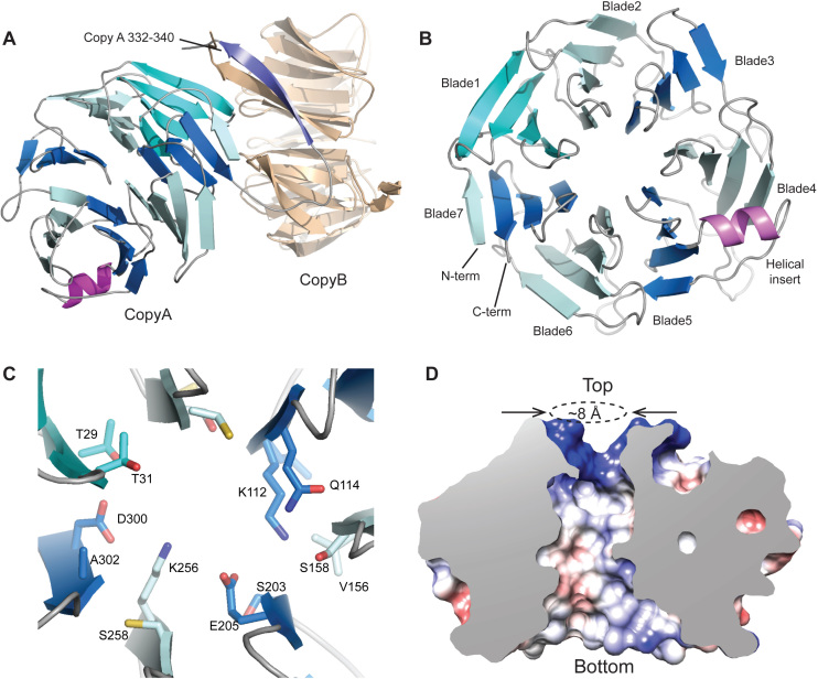
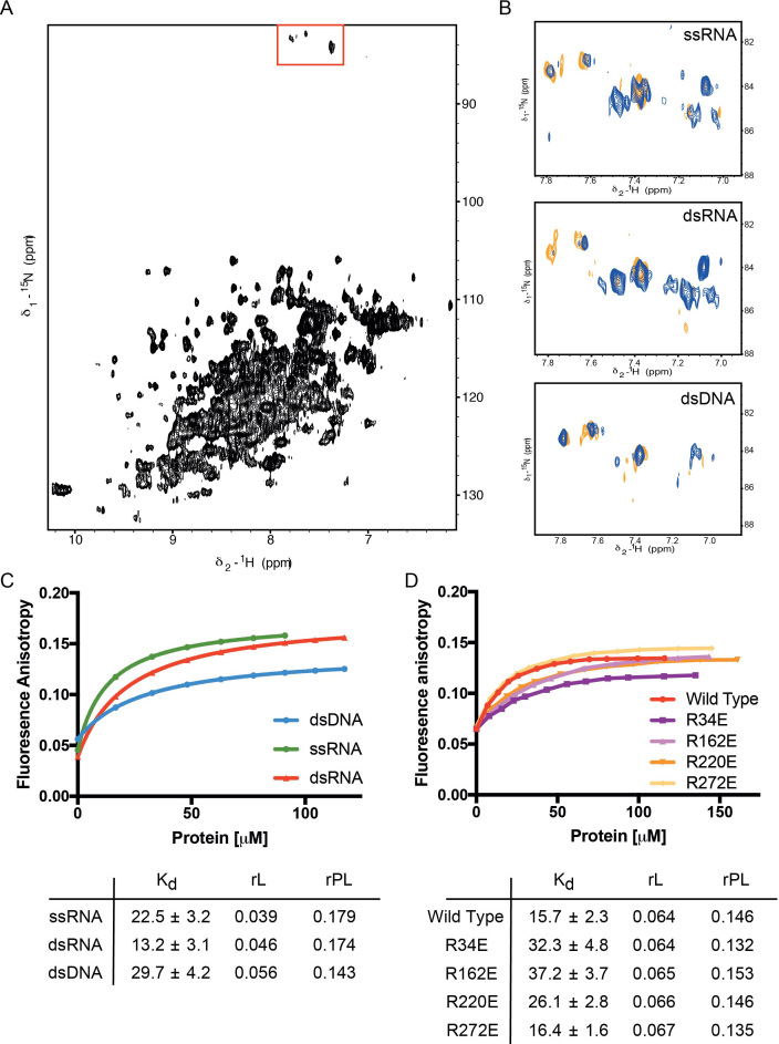
The multi-protein complex WRAD, formed by WDR5, RbBP5, Ash2L and Dpy30, binds to the MLL SET domain to stabilize the catalytically active conformation required for histone H3K4 methylation. In addition, the WRAD complex contributes to the targeting of the activated complex to specific sites on chromatin. RbBP5 is central to MLL catalytic activation, by making critical contacts with the other members of the complex. Interestingly its only major structural domain, a canonical WD40 repeat β-propeller, is not implicated in this function. Here, we present the structure of the RbBP5 β-propeller domain revealing a distinct, feature rich surface, dominated by clusters of Arginine residues. Our nuclear magnetic resonance binding data supports the hypothesis that in addition to the role of RbBP5 in catalytic activation, its β-propeller domain is a platform for the recruitment of the MLL complexes to chromatin targets through its direct interaction with nucleic acids.


Figures

Figure 1.
Figure 1.
RbBP5 architecture and activity. (A) Schematic representation of the RbBP5 protein. The WD40 repeats are indicated along with the three regions shown to interact with the MLL1 SET domain, Ash2L SPRY domain and WDR5. The bars beneath represent the constructs used in the current study. (B) Normalized in vitro methyltransferase assay with H3 peptide substrate for the MLL1 (SET domain) + WDR5 + Ash2L (SPRY domain) with different RbBP5 constructs showing that only the RbBP5 340–380 region is essential for activity. (C) In vitro methyltransferase assay with recombinant mononucleosome substrate (147 bp DNA), using a histone H3K4 monomethyl antibody to follow activity. The loading control gel, stained with Coomassie Blue, is presented in Supplementary Figure S1. (D) Methyltransferase assay with MLL1 (SET domain) + WDR5 + Ash2L (SPRY domain) with RbBP5 construct plus and minus β propeller analysed by MALDI TOF mass spectrometry.
Figure 2.
Figure 2.
Structure of the RbBP5 β-propeller domain. (A) Cartoon representation of the RbBP5 asymmetric unit. The blades of the Copy A β-propeller are colored in alternating shades of blue. Copy B is colored in beige. The Copy A residues 332–340 displace the N-terminal strand in Copy B to form a crystallographic dimer. (B) Top view of the RbBP5 β-propeller showing a canonical arrangement of seven 4-stranded blades. A helical insert (pink) is located at the interface of blades 4 and 5. (C) The RbBP5 central channel is formed from largely polar residues. (D) Cut-through of RbBP5 (electrostatic surface representation) revealing the wide polar channel.
Figure 3.
Figure 3.
Surface features of RbBP5. (A, C and E) Views of the ‘top’ of the RbBP5 β-propeller, showing (A) Electrostatic surface of the ‘top’ face of the β-propeller. The position of the helical insert is indicated. (C) Cartoon representation of the ‘top’ face of the β-propeller. The Arginine (magenta) and Lysine (purple) side chains are shown, with the Arginine ring feature indicated by a blue ring. (E) Sequence conservation mapped onto the top face of the β-propeller (Gradient: green (conserved) to magenta (variable)). (B, D and F) Views of the ‘bottom’ surface of the propeller; (B) Electrostatic surface of the ‘bottom’ face of the β-propeller. (D) Bottom view of the propeller showing the Arginine (magenta) and lysine (purple) side chains. (F) Sequence conservation mapped onto the ‘bottom’ face of the β-propeller.
Figure 4.
Figure 4.
Analysis of RNA binding to the RbBP5 β-propeller. (A) 1H15N HSQC spectrum of the RbBP51–340 construct showing well-dispersed resonances. (B) Titration of a randomized RNA 7-mer oligo nucleotide induces selective resonance shifts in the 1H15N HSQC spectrum. Blue apo RbBP5, orange 1:1, red 4:1 RNA:protein. (C) Scaffold Independent Analysis showing the preference of RbBP5 for each nucleobase at five consecutive positions in the oligonucleotide. (D) Titration of a sequence optimised RNA 7-mer induces selective resonance shifts in the1H15N HSQC NMRspectrum of RbBP5.
Figure 5.
Figure 5.
RbBP5 binding to nucleic acids. (A) 1H15N HSQC spectrum of RbBP51–340 construct optimized for the detection of arginine side chain moieties, indicated by red box at top. (B) Spectrum of RbBP5 free and titrated with ssRNA, dsRNA and dsDNA (orange apo RbBP5, blue 4:1 NA:protein). (C) Fluorescence anisotropy measurements of RbBP51–340 binding to fluorescein labeled nucleic acid. (D) Fluorescence anisotropy measurements of acidic mutants of RbBP51–340 binding to fluorescein labeled dsRNA.

Similar articles

Cited by

References

    1. Berger S.L. The complex language of chromatin regulation during transcription. Nature. 2007; 447:407–412. - PubMed
    1. Kouzarides T. Chromatin modifications and their function. Cell. 2007; 128:693–705. - PubMed
    1. Schuettengruber B., Chourrout D., Vervoort M., Leblanc B., Cavalli G.. Genome regulation by polycomb and trithorax proteins. Cell. 2007; 128:735–745. - PubMed
    1. Bannister A.J., Kouzarides T.. Regulation of chromatin by histone modifications. Cell Res. 2011; 21:381–395. - PMC - PubMed
    1. Ringrose L., Paro R.. Epigenetic regulation of cellular memory by the Polycomb and Trithorax group proteins. Annu. Rev. Genet. 2004; 38:413–443. - PubMed

Publication types

MeSH terms