. 2018 Jun 13;13(6):e0198523.
doi: 10.1371/journal.pone.0198523. eCollection 2018.

Recombinant purified buffalo leukemia inhibitory factor plays an inhibitory role in cell growth

Affiliations

Recombinant purified buffalo leukemia inhibitory factor plays an inhibitory role in cell growth

Syed Azmal Ali et al. PLoS One. .

Abstract

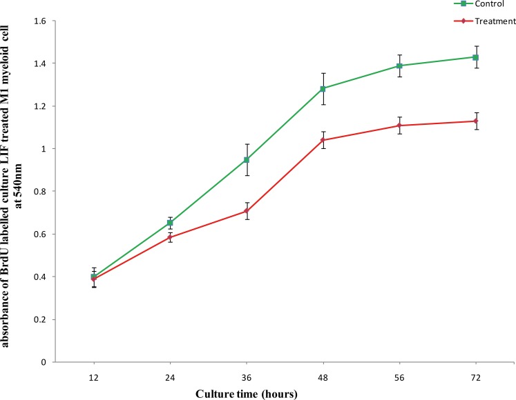
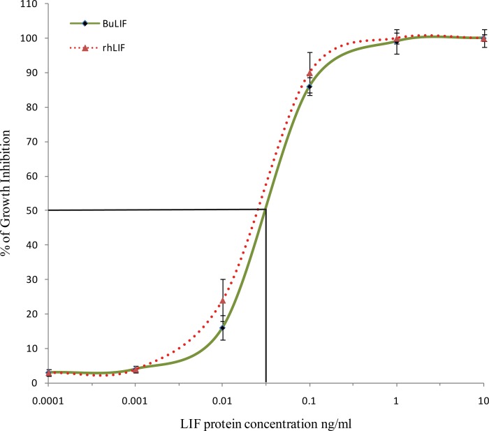
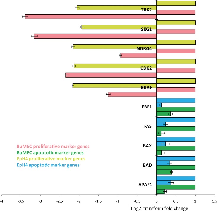
Leukemia Inhibitory Factor (LIF) is a polyfunctional cytokine, involved in numerous regulatory effects in vivo and in vitro, varying from cell proliferation to differentiation, and has therapeutic potential for treating various diseases. In the current study, a COS-1 cell line overexpressing recombinant Buffalo LIF (rBuLIF) was established. The rBuLIF was purified to homogeneity from the total cell lysate of COS-1 cells using a two-step affinity chromatography. The purified LIF was confirmed by western blot and mass spectrometer (MS/MS). Particularly, high-resolution MS has identified the rBuLIF with 73% of sequence coverage with highest confidence parameters and with the search engine score of 4580. We determined the molecular weight of rBuLIF protein to be 58.99 kDa and 48.9 kDa with and without glycosylation, respectively. Moreover, the purified rBuLIF was verified to be functionally active by measuring the growth inhibition of M1 myeloid leukemia cells, revealing a maximum inhibition at 72 hours and half-maximal effective concentration (EC50) of 0.0555 ng/ml, corresponding to a specific activity of >1.6×10(7) units/mg. Next, we evaluated the effect of rBuLIF on buffalo mammary epithelial cell lines for its role in involution and also identified the IC50 value for BuMEC migrating cells to be 77.8 ng/ml. Additionally, the treatment of MECs (BuMEC and EpH4) displayed significant (P < 0.05) reduction in growth progression, as confirmed by qRT-PCR analysis, suggesting its strong involvement in the involution of the mammary gland in vivo. Thus, we conclude that the glycosylated rBuLIF, purified from COS-1 cells was found to be functionally active as its natural counterpart.


Conflict of interest statement

The authors have declared that no competing interests exist.

Figures

Fig 1
Fig 1. Expression of LIF in COS-1 cells.
(A) GFP-tagged full-length LIF was expressed in COS-1 cells and detected by western blot as a secretory protein in the media as well as in total cell lysate. (B) SDS-PAGE Profile: Gel showing the initial washing fractions of the rBuLIF protein during affinity purification of whole cell lysate from the first run of purification. (C) Initially, collected fractions during the chromatographic run from Lane 1, 2 and 3 all the proteins obtained were concentrated in a lyophilizer 1:20 folds to visualize clearly in the SDS gel. M lane for the marker, first run elution profile of the peak shown in the Fig 2A-lanes 4–8. (D) The second run of the chromatographic elution: Eluted fractions from the first run were subjected to the same packed column for the separation of the proteins, based on the affinity. (E) Western blot image of purified rBuLIF protein for confirmation. M lane for the marker, PT lane for PNGase F treated BuLIF without glycan moieties showing 10 kDa reduction in size, PN lane for non-PNGase treated showing intact purified rBuLIF protein of 58.9 kDa molecular weight.
Fig 2
Fig 2. Chromatograms of rBuLIF-GFP protein during affinity purification using Anti-GFP antibodies from whole cell lysate.
(A) First run of chromatography for purification (SDS profile shown in Fig 1B and 1C). (B) The second run of the purification, sample obtained from the first run peak was injected into the column for the second round of purification (respective SDS profile is shown in Fig 1D). The blue line in the chromatogram indicates the elution profile of the proteins while the green line shows the gradient applied.
Fig 3
Fig 3. Cell proliferation assay of active purified rBuLIF on the timescale.
The inhibition of growth of murine myeloid leukaemia M1 cells was measured by BrdU assay on different time points. The error bars indicate the standard deviation.
Fig 4
Fig 4. Bioactivity assay of rBuLIF on murine myeloid leukaemia M1 cells by BrdU assay.
Comparison of growth inhibition of M1 cells incubated with purified rBuLIF (solid line) and commercial rhLIF (dotted line).
Fig 5
Fig 5. Cell migration inhibitory effect of rBuLIF on buffalo mammary epithelial cells.
(A) Cells were induced by addition of EGF (50 ng/mL) as a stimulant for migrating cells in migration assay that increase the migration rate approx. 2.421-fold. In the presence of rBuLIF protein (100 ng/mL), the EGF induced migration of the BuMEC cells was completely abolished. (B) Determination of IC50 value for the cell migration inhibition effect of rBuLIF. Increasing concentrations of rBuLIF protein were used in the cell migration assay which was plotted against the number of migrated cells and the best fit inflexion point was determined for the IC50 value of rBuLIF.
Fig 6
Fig 6. Treatment of rBuLIF to mammary epithelial cells.
(A) Mice mammary epithelial cells–EpH4 without rBuLIF treatment as a control. (B) EpH4 cells treated with purified rBuLIF exhibit changes in morphology forming globule structures. (C) EpH4 cells were treated with commercially available rhLIF. (D) Normal buffalo mammary epithelial cells (BuMEC) in full confluency under bright light. (E) BuMEC cells treated with rBuLIF also exhibit changes in morphology forming globule structures. (F) BuMEC cells treated with rhLIF also showed changes in morphology used as a positive control under bright light. Yellow arrows indicate the globular structures. Scale bar, 500 μm and 10X magnification.
Fig 7
Fig 7. Real-time PCR analysis of proliferative and apoptotic marker genes in rBuLIF treated mammary epithelial cells.
Proliferative and apoptotic marker genes were analyzed after treatment of rBuLIF on BuMEC and EpH4 cells. Fold changes were represented in the form of log2 transform values.

Similar articles

Cited by

References

    1. Taga T. gp130 and the interleukin-6 family cytokines. Annu. Rev. Immunol. 1997, 15:797–819. doi: 10.1146/annurev.immunol.15.1.797 - DOI - PubMed
    1. Tomida M, Yamamoto-Yamaguchi Y, Hozumi M. Characterization of a factor inducing differentiation of mouse myeloid leukemic cells purified from conditioned medium of mouse Ehrlich ascites tumor cells. FEBS Lett. 1984, 178:291–6. - PubMed
    1. Gearing D, Gough N, King J, Hilton D, Nicola N, Simpson R, et al. Molecular cloning and expression of cDNA encoding murine myeloid leukemia inhibitory factor (LIF). EMBO J. 1987, 6:3995–4002. - PMC - PubMed
    1. Smith AG, Heath JK, Donaldson DD, Wong GG, Moreau J, Stahl M, et al. Inhibition of pluripotential embryonic stem cell differentiation by purified polypeptides. Nature. 1988, 336:688–690. doi: 10.1038/336688a0 - DOI - PubMed
    1. Smith A. Embryonic Stem Cells In Stem Cell Biology, Marshak D.R., Gardner R.L., and Gottlieb D., eds. (Cold Spring Harbor, NY: Cold Spring Harbor Laboratory Press; ), 2001, pp. 205–230.

Publication types

Substances

LinkOut - more resources