. 2018 Jun 14;18(1):13.
doi: 10.1186/s12861-018-0172-6.

Developmental nicotine exposure affects larval brain size and the adult dopaminergic system of Drosophila melanogaster

Affiliations

Developmental nicotine exposure affects larval brain size and the adult dopaminergic system of Drosophila melanogaster

Melanie Morris et al. BMC Dev Biol. .

Abstract

Background: Pregnant women may be exposed to nicotine if they smoke or use tobacco products, nicotine replacement therapy, or via e-cigarettes. Prenatal nicotine exposure has been shown to have deleterious effects on the nervous system in mammals including changes in brain size and in the dopaminergic system. The genetic and molecular mechanisms for these changes are not well understood. A Drosophila melanogaster model for these effects of nicotine exposure could contribute to faster identification of genes and molecular pathways underlying these effects. The purpose of this study was to determine if developmental nicotine exposure affects the nervous system of Drosophila melanogaster, focusing on changes to brain size and the dopaminergic system at two developmental stages.

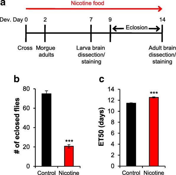
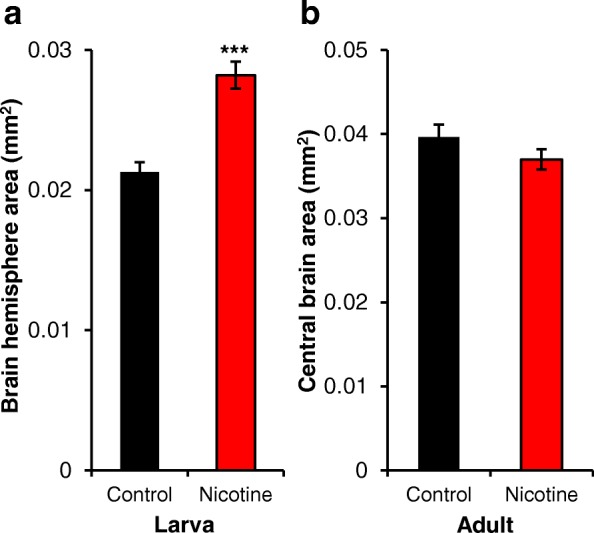
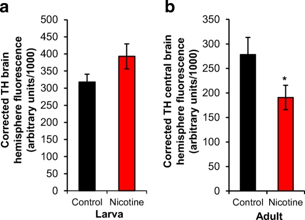
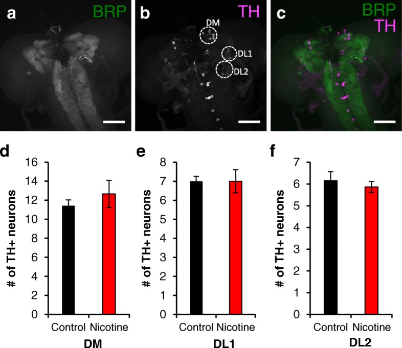
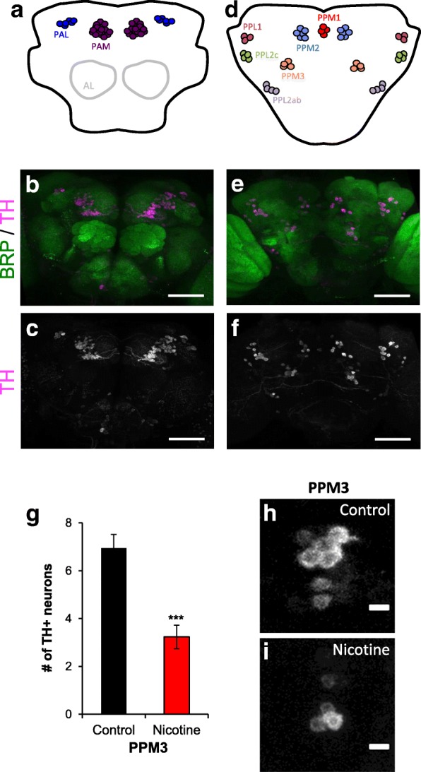
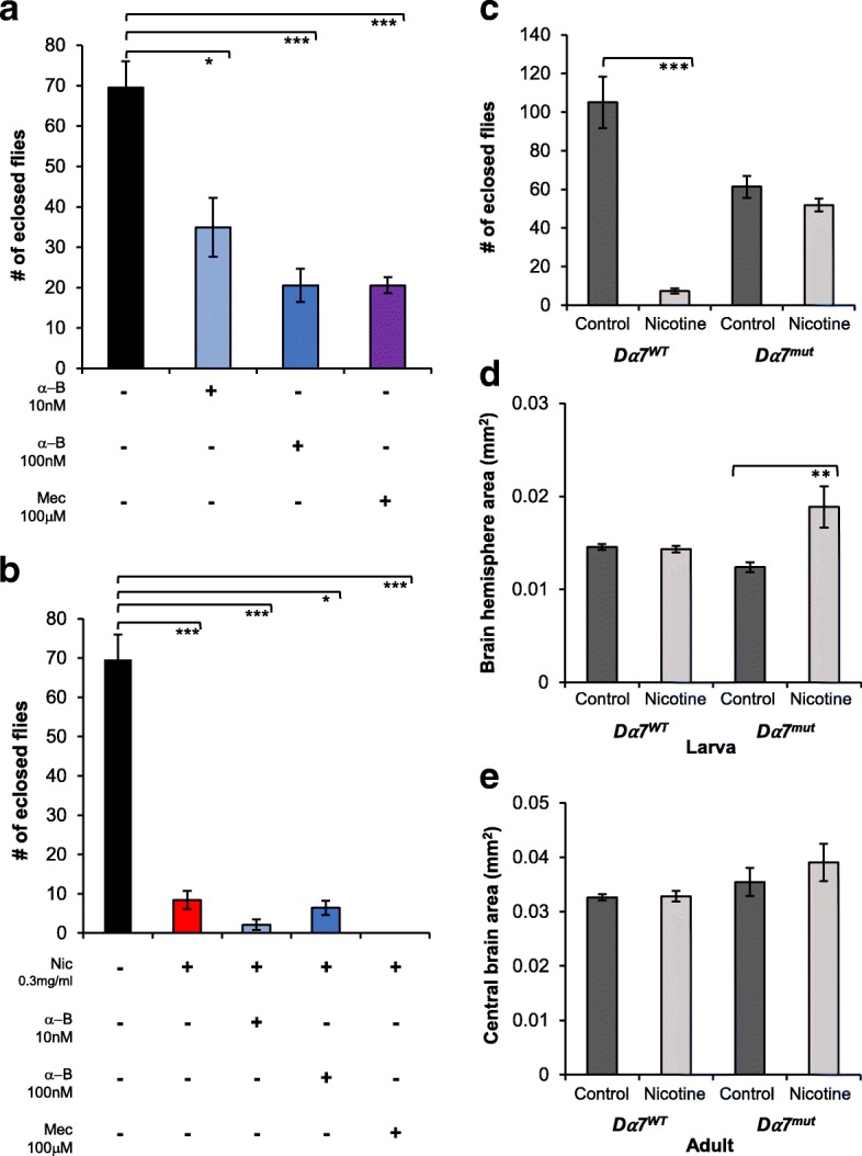
Results: We reared flies on control or nicotine food from egg to 3rd instar larvae or from egg to adult and determined effectiveness of the nicotine treatment. We used immunohistochemistry to visualize the whole brain and dopaminergic neurons, using tyrosine hydroxylase as the marker. We measured brain area, tyrosine hydroxylase fluorescence, and counted the number of dopaminergic neurons in brain clusters. We detected an increase in larval brain hemisphere area, a decrease in tyrosine hydroxylase fluorescence in adult central brains, and a decrease in the number of neurons in the PPM3 adult dopaminergic cluster. We tested involvement of Dα7, one of the nicotinic acetylcholine receptor subunits, and found it was involved in eclosion, as previously described, but not involved in brain size.

Conclusions: We conclude that developmental nicotine exposure in Drosophila melanogaster affects brain size and the dopaminergic system. Prenatal nicotine exposure in mammals has also been shown to have effects on brain size and in the dopaminergic system. This study further establishes Drosophila melanogaster as model organism to study the effects of developmental nicotine exposure. The genetic and molecular tools available for Drosophila research will allow elucidation of the mechanisms underlying the effects of nicotine exposure during development.

Keywords: Brain; Development; Dopamine; Drosophila; Dα7; Nicotine.


Conflict of interest statement

Ethics approval and consent to participate

Drosophila melanogaster is not currently regulated by the Institutional Animal Care and Use Committee (IACUC) of Lewis & Clark College, and hence approval was not seeked. Disposal of Drosophila melanogaster is supervised by the Institutional Biosafety Committee at Lewis & Clark College and this project was carried out according to the approved protocol (IBC-14-01).

Competing interests

The authors declare that they have no competing interests.

Publisher’s Note

Springer Nature remains neutral with regard to jurisdictional claims in published maps and institutional affiliations.

Figures

Fig. 1
Fig. 1
Developmental nicotine treatment affects survival and development time. a Schematic showing the experimental design for the nicotine treatment and for when dissections were carried out. Flies were reared on control food (black bars) or food laced with 0.3 mg/ml nicotine (red bars) and the number of flies eclosed was counted from days 9 to 14 after egg laying to estimate survival and the time required for 50% of pupae to eclose (ET50). b The number of eclosed flies by day 14 was significantly reduced by the nicotine treatment. c The number of days needed for 50% of the flies to eclose was significantly increased by the nicotine treatment. b, c Samples size was n = 64 vials counted for control and n = 95 vials for nicotine from 17 independent experiments. Mann-Whitney U-test (a) and Student’s t-test (b) were used to compare the control versus the nicotine condition
Fig. 2
Fig. 2
Developmental nicotine treatment increases larval brain hemisphere area. Flies were raised on control food (black bars) or food laced with 0.3 mg/ml nicotine (red bars). Larvae were dissected at the 3rd instar stage of development; adults were dissected 4 days after eclosion. The brains were stained with anti-bruchpilot for visualization. a Larval brain hemisphere area was significantly larger in brains from nicotine-exposed larvae. b Adult brains had no difference in central brain area between conditions. Sample size for the larval stage was n = 35 brain hemispheres for control and n = 28 brain hemispheres for nicotine from 6 independent experiments and for adult was n = 24 brains for control and n = 21 brains for nicotine from 9 independent experiments. Student’s t-test was used to compare the control versus the nicotine condition
Fig. 3
Fig. 3
Developmental nicotine exposure decreases TH fluorescence in adult brains. Flies were raised on control food or food laced with 0.3 mg/ml nicotine. Larvae were dissected at the 3rd instar stage of development; adults were dissected 4 days after eclosion. The brains were stained with an antibody against tyrosine hydroxylase (TH). a Corrected TH brain hemisphere fluorescence, which normalized the staining fluorescence to background levels, showed that developmental nicotine exposure had no statistically significant effect on TH staining in larval brains. b Corrected TH central brain fluorescence was significantly decreased in adult brains of flies exposed to nicotine during development. Sample size was n = 22 brain hemispheres for control and n = 18 brain hemispheres for nicotine from 5 independent experiments for the larval stage, and n = 10 brains for control and n = 17 for nicotine from 5 independent experiments for adult. Mann-Whitney U-test (a) and Student’s t-test (b) were used to compare the control versus the nicotine condition
Fig. 4
Fig. 4
Developmental nicotine exposure does not alter the number of TH+ neurons in larval brains. 3rd instar larvae brains were dissected, immunostained, mounted and imaged on a confocal microscope. a-c are maximum projection images of a brain from a larva reared in control food and show representative images of larval brains used for analysis. a An anti-bruchpilot (BRP) antibody was used as background staining for the whole brain. b An anti-tyrosine hydroxylase (TH) antibody was used as marker for dopaminergic neurons. c Merged image of the BRP and TH channels. The scale bar is 50 μM. d-f show the average number of TH+ neurons counted in the DM, DL1 and DL2 dopaminergic clusters from larvae reared in control food (black bars) or nicotine food (red bars). The number of TH+ neurons was not affected by developmental nicotine exposure. Sample size was n = 19 brain hemispheres for control and n = 15 brain hemispheres for nicotine from n = 4 independent experiments. Student’s t-test was used to compare the control versus the nicotine condition
Fig. 5
Fig. 5
Developmental nicotine exposure decreases the number of TH+ neurons in the PPM3 cluster. Flies were reared on control or 0.3 mg/ml nicotine food. Adult brains were dissected, immunostained, mounted and imaged on a confocal microscope. a, d Schematic of the approximate position of the adult dopaminergic clusters. a Anterior clusters shown, d posterior clusters shown. b, c Maximum projection images of the anterior region of a brain from an adult fly that was reared in control food. The PAM and PAL dopaminergic clusters are visible. e, f are maximum projection images of the posterior region of the same brain. The PPM1, PPM2, PPM3, PPL1, PPL2ab and PPL2c are visible. c, f Anti-tyrosine hydroxylase (TH) was used as marker for dopaminergic neurons. b, e Anti-bruchpilot (BRP) was used as background staining for the whole brain. Merged images of the BRP and TH channels. Scale bar 50 μM. g Average number of TH+ neurons counted in the PPM3 cluster of flies reared in control food (black bars) or nicotine food (red bars). h, i Representative images of the PPM3 cluster from flies reared in control (h) or nicotine food (i). The number of TH+ neurons in the PPM3 cluster was reduced in brains from nicotine-exposed flies. Sample size was n = 20 brain hemispheres for control and n = 34 brain hemispheres for nicotine from n ≥ 5 independent experiments. Student’s t-test was used to compare the control versus the nicotine condition
Fig. 6
Fig. 6
Dα7 mediates the effect of nicotine on eclosion but not on larval brain size. a, b wB flies were reared on control (black bars) or 0.3 mg/ml nicotine (red bars) food and the number of eclosed flies was determined. a wB flies were reared in control food or food with nAChRs blockers: 10 nM (light blue bars) or 100 nM (dark blue bars) α-bungarotoxin (α-B) or 100μM (purple bars) mecamylamine (Mec); these drugs significantly decreased eclosion. b Blocking nAChRs during developmental nicotine did not revert the effect of nicotine. c-e Dα7WT (dark grey bars) or Dα7mut (light grey bars) flies were reared on control or 0.1 mg/ml nicotine food and the number of eclosed flies or brain size was determined. c Dα7 mediates the effects of developmental nicotine exposure on eclosion. d, e Dα7 does not regulate larval or adult brain size. a, b wB flies. Sample size: n = 14 vials, control food; n = 18, 0.3 mg/ml nicotine food; n = 12 vials each for 10 nM or 100 nM α-bungarotoxin, 100μM mecamylamine,100 nM α-bungarotoxin+ 0.3 mg/ml nicotine, 100μM mecamylamine + 0.3 mg/ml nicotine; n = 10 vials, 10 nM α-bungarotoxin+ 0.3 mg/ml nicotine; 2 independent experiments for nAChR blockers, 3 for control and nicotine food. c Sample size: n = 12 vials for Dα7WT and Dα7mut, control food; n = 18, Dα7WT nicotine food; n = 16, Dα7mut nicotine food from 3 independent experiments. d Sample size: n = 10 larval brain hemispheres Dα7WT, control food; n = 10, 0.1/mg/ml nicotine food; n = 6 Dα7mut, control food; n = 11, 0.1 mg/ml nicotine food from 3 independent experiments, except Dα7mut in control food, single experiment. e Sample size: n = 6 brains, Dα7WT control food; n = 16, 0.1/mg/ml nicotine food; n = 7 Dα7mut, control food; n = 4, 0.1 mg/ml nicotine food from 2 independent experiments. a-e Kruskal-Wallis test, followed by pairwise comparisons adjusted for multiple comparisons

References

    1. U.S. Department of Health and Human Services. The health consequences of smoking: 50 years of progress. A report of the surgeon general. U.S. Department of Health and Human Services, Centers for Disease Control and PRevention, National Center for Chronic Disease Prevention and Health Promotion, Office on Smoking and Health; 2014. https://www.surgeongeneral.gov/library/reports/50-years-of-progress/full....
    1. World Health Organization. Tobacco: Fact sheet. 2017. http://www.wpro.who.int/mediacentre/factsheets/fs_201203_tobacco/en/.
    1. Center for Behavioral Health Statistics and Quality, Substance Abuse and Mental Health Services Administration, U.S. Department of Health and Human Services, RTI International. Substance and Mental Health Services Administration, Results from the 2013 National Survey on Drug Use and Health: Summary of National Findings. Substance Abuse and Mental Health Services Administration; 2014. https://store.samhsa.gov/product/Results-from-the-2013-National-Survey-o....
    1. Slotkin TA. Cholinergic systems in brain development and disruption by neurotoxicants: nicotine, environmental tobacco smoke, organophosphates. Toxicol Appl Pharmacol. 2004;198:132–151. doi: 10.1016/j.taap.2003.06.001. - DOI - PubMed
    1. Pauly JR, Slotkin TA. Maternal tobacco smoking, nicotine replacement and neurobehavioural development. Acta Paediatr. 2008;97:1331–1337. doi: 10.1111/j.1651-2227.2008.00852.x. - DOI - PubMed

Publication types

MeSH terms

LinkOut - more resources