. 2018 Jun 14;16(1):30.
doi: 10.1186/s12964-018-0244-z.

MicroRNA-644a promotes apoptosis of hepatocellular carcinoma cells by downregulating the expression of heat shock factor 1

Affiliations

MicroRNA-644a promotes apoptosis of hepatocellular carcinoma cells by downregulating the expression of heat shock factor 1

Wenjin Liang et al. Cell Commun Signal. .

Erratum in

Abstract

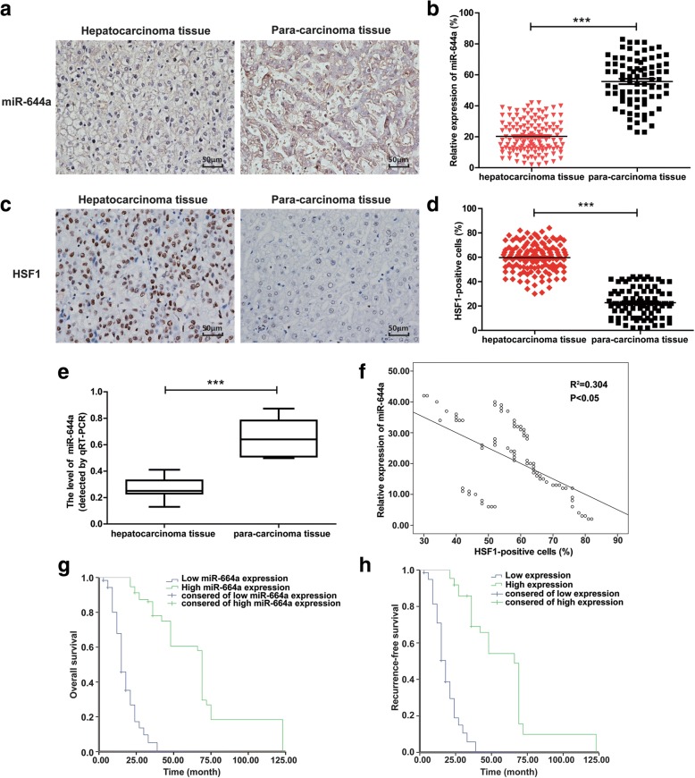
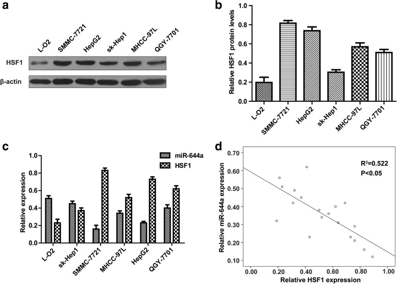
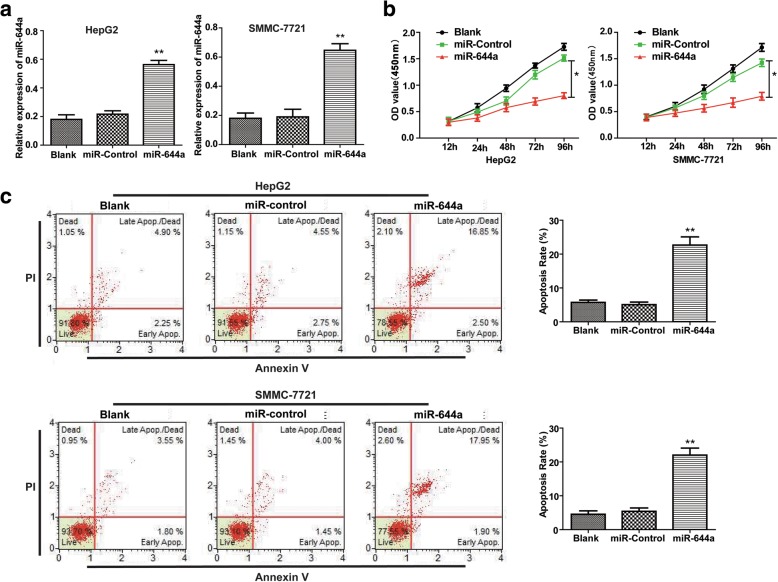
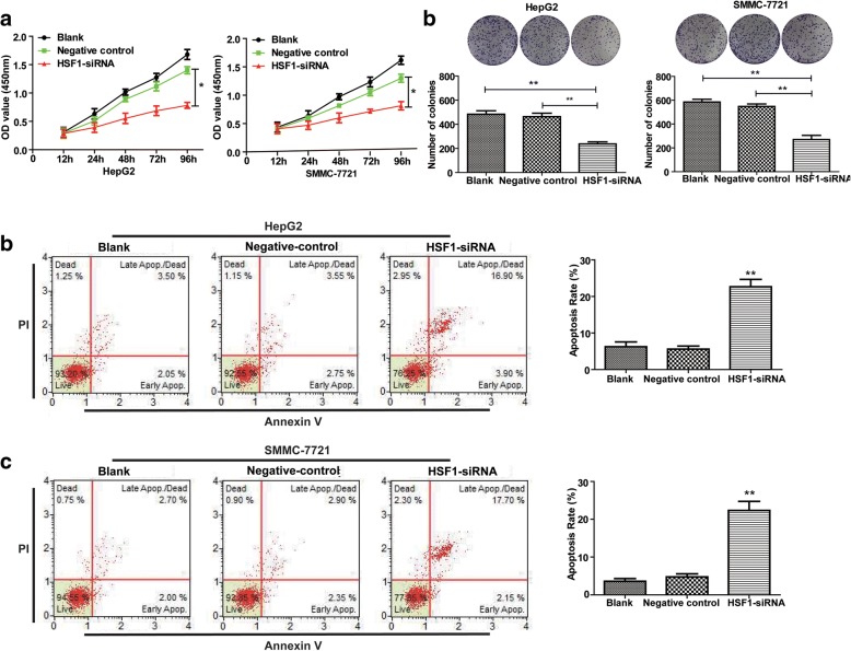
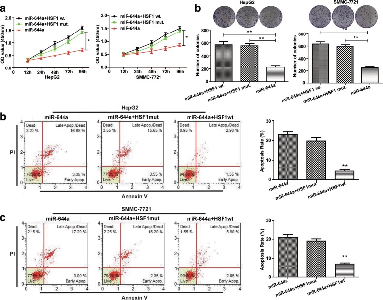
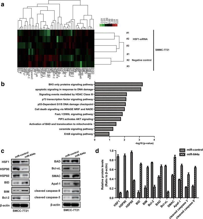
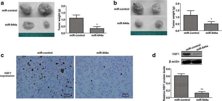
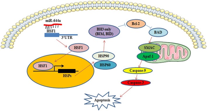
In this study, we investigated the role of microRNA-644a (miR-644a) in the growth and survival of hepatocellular carcinoma (HCC) cells. MiR-644a levels were lower in HCC tissues than in adjacent peri-cancerous tissues (n = 135). MiR-644a expression was inversely correlated with heat shock factor 1 (HSF1) expression, tumour diameter and TNM stage. Moreover, HepG2 and SMMC-7721 cell lines showed lower miR-644a expression than normal L-O2 hepatocytes. MiR-644a overexpression in HepG2 and SMMC-7721 cells increased apoptosis by downregulating HSF1. Dual luciferase reporter assays confirmed the presence of a miR-644a binding site in the 3'-untranslated region (3'-UTR) of HSF1. Xenograft tumours derived from SMMC-7721 cells transfected with a miR-664a mimic showed less growth than tumours derived from untransfected controls. Protein chip analysis revealed that miR-644a-overexpressing SMMC-7721 and HepG2 cells strongly expressed pro-apoptotic BH3-only proteins, such as BID, BAD, BIM, SMAC, Apaf-1 and cleaved caspases-3 and -9. These findings suggest miR-644a promotes apoptosis in HCC cells by inhibiting HSF1.

Keywords: Apoptosis; BH3-only protein; HSF1; Hepatocellular carcinoma; miR-644a.


Conflict of interest statement

Ethics approval and consent to participate

Clinical data have been approved by the Ethics Committee of Guilin Medical.

University and approved by the patients. All animal experiments were approved by Animal Care and Use Committee of Guilin Medical University.

Competing interests

No potential conflicts of interest were disclosed by all authors.

Publisher’s Note

Springer Nature remains neutral with regard to jurisdictional claims in published maps and institutional affiliations.

Figures

Fig. 1
Fig. 1
Low expression of miR-644a in HCC tissues negatively correlates with HSF1 expression and a poor prognosis. (a) Representative images (× 400) show in situ hybridization (ISH) of miR-644a in HCC and adjacent peri-cancerous liver tissues. (b) Quantitative analysis of miR-644a expression in HCC and adjacent peri-cancerous liver tissues (n = 135/group) based on ISH. Note: *** denotes P < 0.001 when compared to adjacent tissues. (c) Representative IHC images (× 400) show HSF1 expression in HCC and adjacent peri-cancerous tissues (n = 135/group). (d) Quantitative analysis of HSF1 expression in HCC and adjacent peri-cancerous liver tissues (n = 135/group) based on IHC. Note: *** denotes P < 0.001 when compared to adjacent tissues. (e) Quantitative RT-PCR analysis of miR-644a expression in HCC and adjacent peri-cancerous liver tissues (n = 135/group). Note: *** denotes P < 0.001 when compared to adjacent tissues. (f) Spearman’s test results show low miR-644a expression that negatively correlates with high HSF1 expression in HCC tissues (R2 = 0.304, P < 0.05). (g-h) Kaplan-Meier survival analysis demonstrates a correlation between miR-644a expression in HCC patients and their overall survival (g) and recurrence-free survival (h)
Fig. 2
Fig. 2
Correlation between miR-644a and HSF1 expression in HCC cells. (a) Quantitative RT-PCR analysis of miR-644a expression in HCC cell lines, HepG2, SMMC-7721, sk-Hep1, MHCC-97 L, QGY-7701 and the normal liver cells, L-O2. (b) Representative western blot shows HSF1 protein expression in the HCC cell lines HepG2, SMMC-7721, sk-Hep1, MHCC-97 L, and QGY-7701 and the normal liver cells, L-O2. (c) Quantitative RT-PCR analysis of HSF1 mRNA expression in the HCC cell lines, HepG2, SMMC-7721, sk-Hep1, MHCC-97 L, and QGY-7701 and the normal liver cells, L-O2. (d) Spearman’s analysis shows a correlation between miR-644a and HSF1 expression in the HCC cell lines (R2 = 0.522, P < 0.05). Note: Data are represented as the mean ± SD of three independent experiments and were analysed by a paired t-test
Fig. 3
Fig. 3
MiR-644a upregulation inhibits proliferation and promotes apoptosis of HCC cells. (a) Quantitative RT-PCR analysis of miR-644a expression in control and miR-644a mimic-transfected HepG2 and SMMC-7721 cells. (b) The CCK8 assay shows proliferation in the control and miR-644a mimic-transfected HepG2 and SMMC-7721 cells. (c) Flow cytometry analysis of percent apoptosis (AnnexinV+ PI+) in the control and miR-644a mimic-transfected HepG2 and SMMC-7721 cells. Note: * denotes P < 0.05 and ** denotes P < 0.01 compared to controls
Fig. 4
Fig. 4
MiR-644a downregulates HSF1 expression by binding to its 3’UTR. (a) Targetscan and miRanda analysis show putative miR-644a binding sites in the 3’UTR of HSF1. (b) Quantitative RT-PCR analysis of HSF1 mRNA expression in HepG2 and SMMC-7721 cells co-transfected with miR-644a mimic and wild type or mutant HSF1. (c) Representative western blot shows HSF1 protein expression in HepG2 and SMMC-7721 cells co-transfected with miR-644a mimic and wild type or mutant HSF1. (de) Relative luciferase activity in HepG2 and SMMC-7721 cells co-transfected with miR-644a mimic and wild type or mutant HSF1. Note: * denotes P < 0.05 and ** denotes P < 0.01 compared to controls
Fig. 5
Fig. 5
HSF1 silencing decreases proliferation and increases apoptosis of HCC cells. (a) CCK8 assay shows proliferation of HepG2 and SMMC-7721 cells transfected with control or HSF1-siRNA. (b) Colony formation assay shows the total number of colonies formed by HepG2 and SMMC-7721 cells transfected with control or HSF1-siRNA. (c) Flow cytometry analysis shows apoptosis of HepG2 and SMMC-7721 cells transfected with control or HSF1-siRNA. Note: * denotes P < 0.05 and ** denotes P < 0.01 compared to controls
Fig. 6
Fig. 6
HSF1 overexpression promotes proliferation and inhibits apoptosis of HCC cells. (a) CCK8 assay shows proliferation of HepG2 and SMMC-7721 cells co-transfected with miR-644a mimic and wild-type or mutant HSF1 (wild type or mutant 3’UTR). (b) Colony formation assay shows the total number of colonies in HepG2 and SMMC-7721 cells co-transfected with miR-644a mimic and wild-type or mutant HSF1. (c) Flow cytometry analysis of percent apoptosis (AnnexinV+ PI+) in HepG2 and SMMC-7721 cells co-transfected with miR-644a mimic and wild-type or mutant HSF1. Note: * denotes P < 0.05 and ** denotes P < 0.01 compared to controls
Fig. 7
Fig. 7
MiR-644a/HSF1 axis regulates HCC cell apoptosis via the BH3-only protein signalling pathway. (a) Protein chip clustering analysis shows differential protein expression in control and HSF1 siRNA transfected SMMC-7721 cells. (b) Protein chip enrichment analysis shows signalling pathways differentially regulated by silencing HSF1 in SMMC-7721 cells. (cd) Representative western blots show expression of apoptosis-related proteins in SMMC-7721 cells transfected with miR-644a mimic or control miRNA. All experiments were repeated three times
Fig. 8
Fig. 8
MiR-644a overexpression inhibits xenograft HCC tumour growth. (a) Representative images of xenograft tumours from nude mice subcutaneously injected with SMMC-7721 cells stably transfected with miR-644a mimic or control miRNA. (b) Xenograft tumour weights from nude mice subcutaneously injected with SMMC-7721 cells stably transfected with miR-644a mimic or control miRNA. (c) Representative images (400×) of IHC staining of HSF1 in xenograft tumours derived from SMMC-7721 cells stably transfected with miR-644a mimic or control miRNA. (d) Representative western blot shows HSF1 expression in xenograft tumours derived from SMMC-7721 cells stably transfected with miR-644a mimic or control miRNA. Note: * denotes P < 0.05 and ** denotes P < 0.01 compared to controls
Fig. 9
Fig. 9
Schematic diagram of apoptosis regulation in HCC cells by miR-644a

References

    1. Sia D, Villanueva A, Friedman SL, Llovet JM. Liver Cancer cell of origin, molecular class, and effects on patient prognosis. Gastroenterology. 2017;152(4):745–761. doi: 10.1053/j.gastro.2016.11.048. - DOI - PubMed
    1. Siegel RL, Miller KD, Jemal A. Cancer statistics, 2017. CA Cancer J Clin. 2017;67(1):7–30. doi: 10.3322/caac.21387. - DOI - PubMed
    1. Chen W, Zheng R, Baade PD, Zhang S, Zeng H, Bray F, et al. Cancer statistics in China, 2015. CA Cancer J Clin. 2016;66(2):115–132. doi: 10.3322/caac.21338. - DOI - PubMed
    1. Bartel DP. MicroRNAs: genomics, biogenesis, mechanism. and function Cell. 2004;116(2):281–297. - PubMed
    1. Griffiths-Jones S, Grocock RJ, van Dongen S, Bateman A, Enright AJ. miRBase: microRNA sequences, targets and gene nomenclature. Nucleic Acids Res. 2006;34(Database issue):D140–D144. doi: 10.1093/nar/gkj112. - DOI - PMC - PubMed

Publication types

MeSH terms