Review
. 2018 Jun 13;82(3):e00013-18.
doi: 10.1128/MMBR.00013-18. Print 2018 Sep.

Replication Fork Breakage and Restart in Escherichia coli

Affiliations
Review

Replication Fork Breakage and Restart in Escherichia coli

Bénédicte Michel et al. Microbiol Mol Biol Rev. .

Abstract

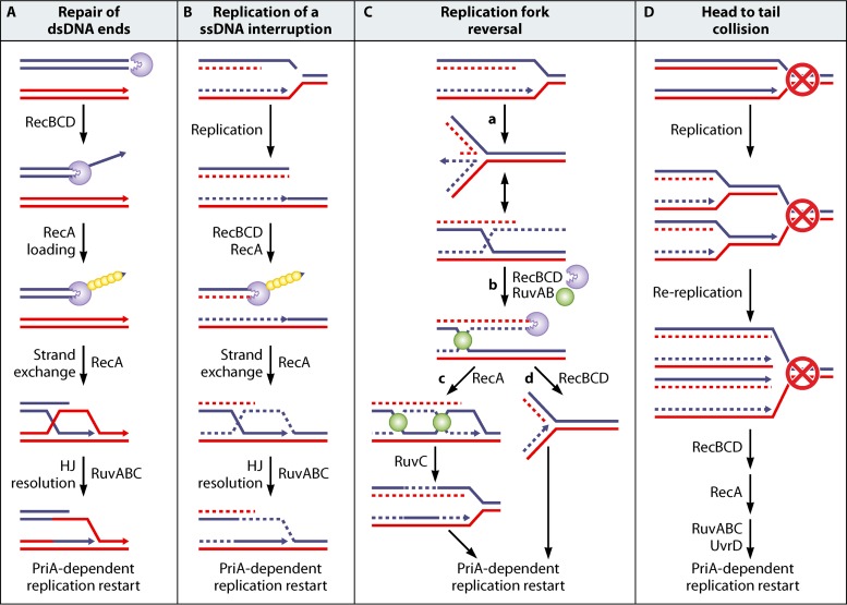
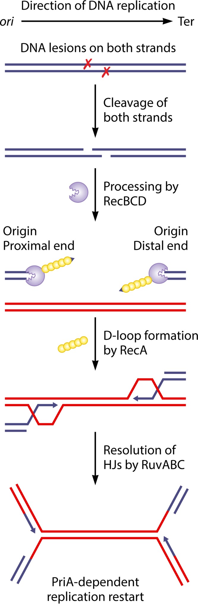
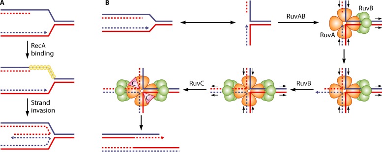
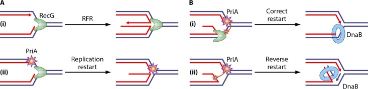
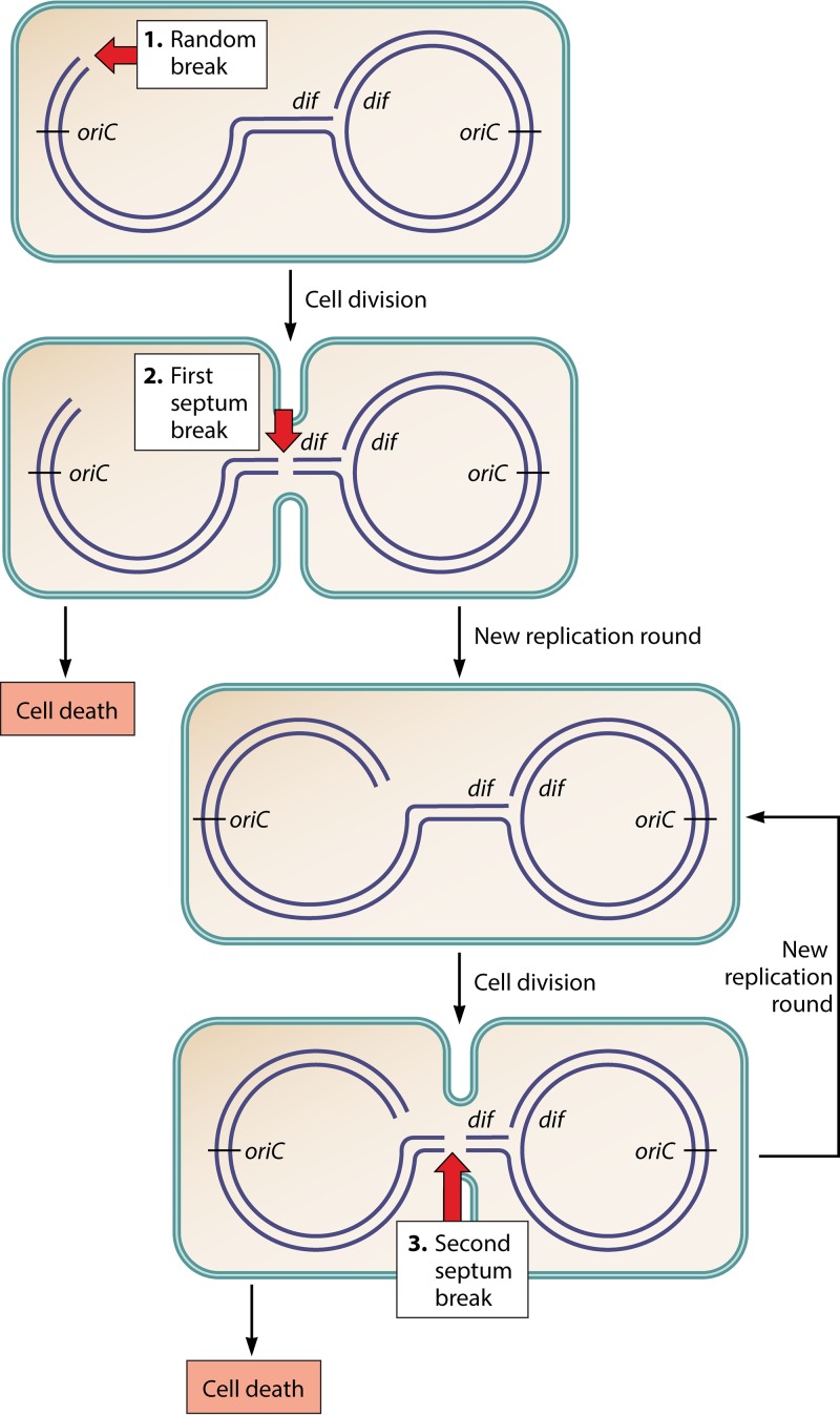
In all organisms, replication impairments are an important source of genome rearrangements, mainly because of the formation of double-stranded DNA (dsDNA) ends at inactivated replication forks. Three reactions for the formation of dsDNA ends at replication forks were originally described for Escherichia coli and became seminal models for all organisms: the encounter of replication forks with preexisting single-stranded DNA (ssDNA) interruptions, replication fork reversal, and head-to-tail collisions of successive replication rounds. Here, we first review the experimental evidence that now allows us to know when, where, and how these three different reactions occur in E. coli. Next, we recall our recent studies showing that in wild-type E. coli, spontaneous replication fork breakage occurs in 18% of cells at each generation. We propose that it results from the replication of preexisting nicks or gaps, since it does not involve replication fork reversal or head-to-tail fork collisions. In the recB mutant, deficient for double-strand break (DSB) repair, fork breakage triggers DSBs in the chromosome terminus during cell division, a reaction that is heritable for several generations. Finally, we recapitulate several observations suggesting that restart from intact inactivated replication forks and restart from recombination intermediates require different sets of enzymatic activities. The finding that 18% of cells suffer replication fork breakage suggests that DNA remains intact at most inactivated forks. Similarly, only 18% of cells need the helicase loader for replication restart, which leads us to speculate that the replicative helicase remains on DNA at intact inactivated replication forks and is reactivated by the replication restart proteins.

Keywords: PriA; RecA; RecBC; RecBCD; RecG; RuvAB; chromosome terminus; double-strand break; recombination; replication fork reversal; replication restart.


Figures

FIG 1
FIG 1
(A) Repair of a broken replication fork by RecBCD, RecA, Ruv, and PriA. RecBCD binds to DNA double-strand ends and degrades both strands until it encounters a Chi site at which it loads RecA onto the 3′ DNA end. The RecA-ssDNA filament invades a homologous region and promotes strand exchange, resulting in a Holliday junction (HJ), indicated by the blue and red crossing lines, and an adjacent displacement loop, also called a D-loop, schematized by the displacement of one of the red lines by the blue line end. RuvAB binding to the HJ drives branch migration. After RuvC binding, the RuvABC complex catalyzes the resolution of the HJ, resulting in a recombinant molecule. PriA restarts replication from the D-loop. (B) Formation of a “broken fork” by the encounter of a preexisting nick. A ssDNA break is drawn here on the lagging-strand template, but the same reaction occurs with a ssDNA break on the leading-strand template. DSB repair is the same as in panel A and reconstitutes a replication fork. (C) Replication fork reversal. In the first step (step a), the replication fork is arrested, and the leading- and lagging-strand ends of the newly synthesized strands anneal. The resulting structure is called a reversed fork; it has a four-arm structure akin to a HJ. Two alternative representations of this structure are shown, called open X and parallel stacked X. RecBC acts on the dsDNA end (as shown in panel A) and is essential for the resetting of the fork, either by RecA-dependent homologous recombination (steps b and c) or by DNA degradation (steps b to d). Either pathway creates a substrate for replication restart proteins (PriA and its partners), since homologous recombination leads to a D-loop, as shown in panel A, and DNA degradation restores a fork structure. In the absence of RecBCD, the resolution of the HJ causes chromosome linearization (not shown). (D) dsDNA ends formed by head-to-tail collision of replication forks. The dsDNA ends formed by rereplication are recombined as in panel A. This reaction occurs at forks blocked at an ectopic Ter site, where it requires UvrD to dislodge the Tus protein (see the text). In panel A, the blue and red continuous double lines represent two homologous DNA molecules. In panels B and C, the continuous lines represent the parental chromosome, and the dashed lines represent the newly synthesized strands. In panel D, the dashed lines represent the DNA synthesized in a second replication round. Arrowheads show DNA 3′ ends. Incised purple circles, RecBCD; small yellow circles, RecA; green circles, RuvAB.
FIG 2
FIG 2
Model for two-ended break repair following the excision of two closely spaced lesions on opposite strands of the DNA. When two lesions (denoted by X) are closely spaced on opposite strands of the DNA, their nucleolytic excision can lead to a two-ended double-strand break behind the replication fork. One of the ends, closest to the origin of replication, is denoted the origin-proximal end, and the other end, closest to the terminus of replication, is denoted the origin-distal end. Each end is processed by the RecBCD enzyme, which loads the RecA protein. The RecA protein catalyzes strand invasion to form two D-loops and two HJs that are resolved by RuvABC. This converts the joint molecules to converging replication forks, which assemble new replisomes through PriA-dependent restart. The blue and red continuous double lines represent two homologous DNA molecules. Arrowheads show DNA 3′ ends.
FIG 3
FIG 3
(A) Model of RFR by RecA. RecA binding to the ssDNA region on the lagging-strand template of a blocked fork can promote the invasion of the homologous sequence on the leading strand. This reaction produces a reversed fork. (B) Model of RFR by RuvAB. The RuvAB complex formed on a replication fork can contain only one RuvB hexamer. Branch migration promoted by this RuvB hexamer extrudes a dsDNA end on which a second RuvB hexamer can bind, resulting in a RuvAB-HJ complex similar to the one formed during homologous recombination. RuvC resolves the HJ, which, in the case of a reversed fork, results in fork breakage. Continuous lines, parental DNA strands; dashed lines, newly synthesized DNA strands; small yellow circles, RecA; orange, trefoil RuvA tetramer; green circles, RuvB. Arrowheads show DNA 3′ ends. The small black arrows indicate the direction of strand displacement by the RuvAB complex.
FIG 4
FIG 4
Model of the concerted action of RecG and PriA at forks. It has been shown that RecG remodels replication forks in vitro. (Ai) When RecG is alone, this remodeling causes RFR. (ii) When both RecG and PriA are present, PriA binds to the 3′ end at the fork, preventing its unwinding by RecG. This reaction is likely to also take place in vivo, since no genetic evidence for RecG-dependent RFR could be obtained. (Bi) It has been proposed that in vivo, in addition to preventing RFR, the binding of PriA in the presence of RecG leads to the correct loading of DnaB to the lagging-strand template. Because the PriA helicase domains, represented as small orange stars, bind to the lagging-strand template, replication restarts in the initial direction. (ii) In the absence of RecG, the PriA helicase domains bind to the newly synthesized lagging strand, and consequently, PriA can load DnaB incorrectly to this strand. This results in reverse restart, the assembly of a replication fork proceeding in the wrong direction. Large blue lines, template strands; red lines, newly synthesized strands; small blue lines in panel Bii, strand synthesized by reverse restart; green crescent, RecG; purple star, PriA; blue ring, DnaB replicative helicase. Arrowheads show DNA 3′ ends.
FIG 5
FIG 5
Model for terminus DNA loss in the recB mutant. In a first step, a replication fork broken at a random position remains unrepaired in a recB mutant, resulting in the inability to complete one chromosome. The two daughter chromosomes, one truncated and one whole, are linked by the intact replication fork and segregate to the two cell halves. In a second step, the terminus region of the truncated chromosome becomes trapped in the septum and is broken at the dif site during cell division. A nonviable cell with a linear chromosome and a viable cell with a sigma-replicating chromosome are generated. In a third step, the intact replication fork on the sigma-replicating chromosome meets a fork coming from the origin, which extends the small tail by the entire chromosome arm. This leads to the same substrate as the one generated originally by the fork breakage event, except that the end of the chromosome arm results from terminus breakage. Breakage of the new terminus DNA will occur again at the next cell division, generating a nonviable cell with a full linear chromosome and a viable cell with a sigma-replicating chromosome, and the same reaction can take place for several generations. (See reference for a more detailed depiction of these events.) Light blue lines, bacteria; dark blue lines, DNA; large red arrows, DNA breaks. The positions of the replication origin oriC and of the last segregated sequence in the terminus, the dif site opposite oriC, are indicated.

Similar articles

Cited by

References

    1. Bierne H, Ehrlich SD, Michel B. 1991. The replication termination signal terB of the Escherichia coli chromosome is a deletion hot spot. EMBO J 10:2699–2705. - PMC - PubMed
    1. Bierne H, Ehrlich SD, Michel B. 1997. Deletions at stalled replication forks occur by two different pathways. EMBO J 16:3332–3340. doi:10.1093/emboj/16.11.3332. - DOI - PMC - PubMed
    1. Vilette D, Uzest M, Ehrlich SD, Michel B. 1992. DNA transcription and repressor binding affect deletion formation in Escherichia coli plasmids. EMBO J 11:3629–3634. - PMC - PubMed
    1. Vilette D, Ehrlich SD, Michel B. 1995. Transcription-induced deletions in Escherichia coli plasmids. Mol Microbiol 17:493–504. doi:10.1111/j.1365-2958.1995.mmi_17030493.x. - DOI - PubMed
    1. Michel B, Ehrlich SD, Uzest M. 1997. DNA double-strand breaks caused by replication arrest. EMBO J 16:430–438. doi:10.1093/emboj/16.2.430. - DOI - PMC - PubMed

Publication types

MeSH terms

LinkOut - more resources