. 2018 Oct 15;24(20):5058-5071.
doi: 10.1158/1078-0432.CCR-17-3427. Epub 2018 Jun 13.

Elective Nodal Irradiation Attenuates the Combinatorial Efficacy of Stereotactic Radiation Therapy and Immunotherapy

Affiliations

Elective Nodal Irradiation Attenuates the Combinatorial Efficacy of Stereotactic Radiation Therapy and Immunotherapy

Ariel E Marciscano et al. Clin Cancer Res. .

Abstract

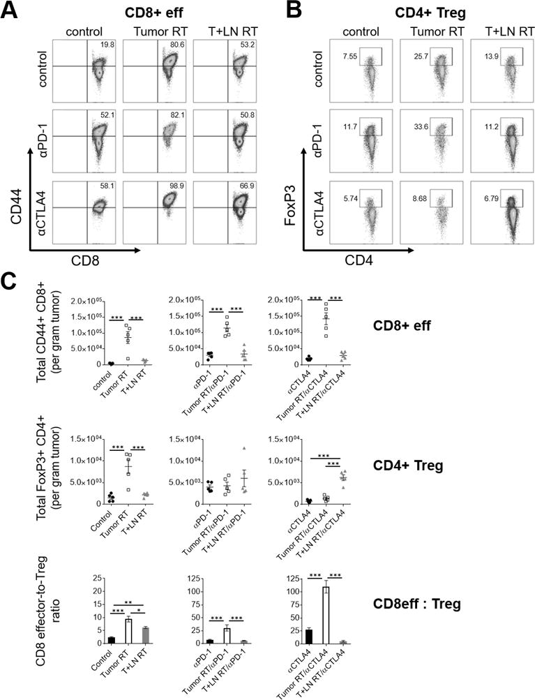
Purpose: In the proper context, radiotherapy can promote antitumor immunity. It is unknown if elective nodal irradiation (ENI), a strategy that irradiates tumor-associated draining lymph nodes (DLN), affects adaptive immune responses and combinatorial efficacy of radiotherapy with immune checkpoint blockade (ICB).Experimental Design: We developed a preclinical model to compare stereotactic radiotherapy (Tumor RT) with or without ENI to examine immunologic differences between radiotherapy techniques that spare or irradiate the DLN.Results: Tumor RT was associated with upregulation of an intratumoral T-cell chemoattractant chemokine signature (CXCR3, CCR5-related) that resulted in robust infiltration of antigen-specific CD8+ effector T cells as well as FoxP3+ regulatory T cells (Tregs). The addition of ENI attenuated chemokine expression, restrained immune infiltration, and adversely affected survival when combined with ICB, especially with anti-CLTA4 therapy. The combination of stereotactic radiotherapy and ICB led to long-term survival in a subset of mice and was associated with favorable CD8 effector-to-Treg ratios and increased intratumoral density of antigen-specific CD8+ T cells. Although radiotherapy technique (Tumor RT vs. ENI) affected initial tumor control and survival, the ability to reject tumor upon rechallenge was partially dependent upon the mechanism of action of ICB; as radiotherapy/anti-CTLA4 was superior to radiotherapy/anti-PD-1.Conclusions: Our results highlight that irradiation of the DLN restrains adaptive immune responses through altered chemokine expression and CD8+ T-cell trafficking. These data have implications for combining radiotherapy and ICB, long-term survival, and induction of immunologic memory. Clinically, the immunomodulatory effect of the radiotherapy strategy should be considered when combining stereotactic radiotherapy with immunotherapy. Clin Cancer Res; 24(20); 5058-71. ©2018 AACR.


Conflict of interest statement

Conflict of Interest Disclosure Statement: CGD is a co-inventor on patents licensed from JHU to BMS. MJS, KBT, and AJK are paid employees of BMS. JW is the inventor of the SARRP and receives royalty and consultancy reimbursement from Xstrahl.

Figures

Figure 1
Figure 1. A preclinical model of image-guided elective nodal irradiation (ENI) accurately targets the tumor-associated DLN
(A) Lymphatic tracking of fluorescent dye (IRDye® 800CW PEG contrast agent) following intratumoral administration on day 11 in tumor-bearing C57BL/6J mice inoculated by s.c. or i.d. flank injection with 5×10⁵ B16F10 or 1.5×10⁶ MC38 cells (n=2-3 mice per experiment, repeated twice). (B) Schematic of intratumoral dye injection, transit and accumulation in ipsilateral tumor-associated inguinal lymph node (DLN). (C) Representative images of fluorescent dye-guided surgery isolating ipsilateral inguinal DLN and coronal view cone-beam CT image with crosshairs overlying inguinal fat pad. (D) Immunofluorescence detection (63X magnification) of phospho-H2AXser139 (green) and DAPI (blue) nuclear staining in non-irradiated (control) and irradiated mice (Tumor RT or T+LN RT). Tumor and DLN tissue harvested 1 hour after 12Gy x1. (E) Representative H&E stain (4-20X magnification) of inguinal DLN harvested from untreated B16F10 tumor-bearing mouse (n=3) on day 14 post-inoculation depicting absence of tumor cells.
Figure 2
Figure 2. Elective nodal irradiation decreases tumor-infiltrating immune cell density relative to tumor-only stereotactic RT
Flank-tumor bearing mice untreated or irradiated (12Gy x1) to tumor-only (Tumor RT) or tumor and DLN (T+LN RT) on day 11 following s.c. injection of 1.5×10⁶ MC38 tumor cells (n=4-9 mice per group, repeated 2-3 times). Tumors were harvested for analysis on day 16, 120 hours after treatment. (A-C) Representative quantitative scatterplot and flow cytometry demonstrating absolute number tumor-infiltrating immune cells and percentage (CD8+ effector, CD44+ CD62L- CD8+; Treg, CD4+ FoxP3+; myeloid derived suppressor cells, CD11b+ Gr-1hi) per gram tumor in MC38 tumor model. (D) Fold change of CD8 effector and Treg subsets by treatment group, normalized to control group. Quantitative bar graph of CD8 effector-to-Treg ratio by treatment group. Error bars represent SEM, ***: p < 0.001, **: p < 0.01, *: p < 0.05, treatment group comparisons by one-way ANOVA and post-hoc Tukey’s multiple comparison test.
Figure 3
Figure 3. Tumor RT and T+LN RT have distinct radiation-induced intratumoral chemokine expression and CD8+ T-cell trafficking patterns
(A) Tumor-associated DLN of MC38 tumor-bearing mice harvested at 1hr, 48hrs and 120hrs after treatment with Tumor RT or T+LN RT on day 11. Quantitative bar graphs representing fold change of absolute number of CD4+, CD8+ and CD11b+ immune cell subsets in tumor-associated DLN over time, normalized to control (n=12 mice per group with 4 mice per time point, repeated twice). (B) Tumor lysate collected at same post-RT timepoints as Fig. 3A and analyzed by Luminex® multiplex immunoassay. Colorimetric heat map conditional formatting with blue to red indicating low to high chemokine/cytokine concentration [pg/mL], respectively. Error bars represent SEM, ***: p < 0.001, **: p < 0.01, *: p < 0.05, CD45+ immune cell and chemokine expression time course experiments analyzed by two-way ANOVA and post-hoc Tukey’s multiple comparison test.
Figure 4
Figure 4. Radiation-mediated tumor infiltration with functional antigen-specific CD8+ T-cells is restrained by elective nodal irradiation
2×10⁶ CFSE-labeled, OVA-specific CD8+ T-cells from donor Rag−/−/OT-1 TCR transgenic CD45.2 mice were adoptively transferred (AT) into MC38-OVA tumor-bearing congenically-mismatched CD45.1 C57BL/6J mice on day 13. CD45.1 mice were treated on day 11 (48 hours before AT) with control, Tumor RT and T+LN RT with DLN and tumor harvested on day 16, 120 hours after RT (AT, n=5-9 mice per group, repeated 3 times). (A) Quantitative scatter plots of effector cytokine production by tumor-infiltrating OT-1, endogenous CD8+ T-cells via intracellular cytokine staining and ELISA (upper rows, L to R respectively). (B) Absolute number of tumor-infiltrating CD8+ CD44+ T-cells per gram tumor by H-2kb OVA (SIINFEKL) tetramer staining and representative flow cytometry plots (ELISA and tetramer staining, n=5 per group, repeated twice). (C) Bar graphs and representative flow plots of effector cytokine production (IFNɣ, TNFα) by AT CD45.2+ CD8+ T-cells in tumor-associated DLN. Error bars represent SEM, ***: p < 0.001, **: p < 0.01, *: p < 0.05, treatment group comparisons by one-way ANOVA and post-hoc Tukey’s multiple comparison test.
Figure 5
Figure 5. Elective nodal irradiation attenuates combinatorial efficacy between radiation and immunotherapy
(A) Treatment schema; Tumor RT or T+LN RT (12Gy x1) administered on day 11 and three doses (i.p. 200μg) of therapeutic antibody (isotype, αPD-1, αCTLA4) on days 10,12,14 in MC38-OVA tumor-bearing mice (n=6-7 per group, repeated twice). (B) Spider plots of tumor outgrowth (mm3) by treatment group, annotated with number of complete responses (CR) over total mice treated. (C) Percent survival by treatment group at day 90 after s.c. flank injection of MC38-OVA tumor cells. (D) Summary table of % CR and median survival by treatment corresponding to (B-C). Kaplan-Meier analysis with log-rank (Mantel-Cox) test for survival differences between treatment groups. ***: p < 0.001, **: p < 0.01, *: p < 0.05.
Figure 6
Figure 6. Favorable modulation of intratumoral CD8 effector-to-Treg ratio is associated with long-term survival
MC38-OVA tumor-bearing mice treated per schema in Fig. 5A, tumors harvested day 21 after s.c. flank injection (n=5 per group). (A-B) Representative flow cytometry (left, CD8 effectors; right, Tregs) and (C) quantitative scatterplots (upper and middle panels), demonstrating percentage and absolute number tumor-infiltrating immune cells per gram tumor by treatment group, respectively. (C) Quantitative bar graph (bottom panel) of CD8 effector-to-Treg ratio by treatment group. Error bars represent SEM, ***: p < 0.001, **: p < 0.01, *: p < 0.05, treatment group comparisons by one-way ANOVA and post-hoc Tukey’s multiple comparison test.
Figure 7
Figure 7. Improved resistance to re-challenge in animals treated with RT + αCTLA4
MC38-OVA tumor-bearing mice treated per schema in Fig. 5A. Tumors harvested day 21 after s.c. flank injection (n=5 per group). (A) Absolute number of antigen-specific tumor-infiltrating CD8+ CD44+ T-cells per gram tumor by H-2kb OVA (SIINFEKL) tetramer staining. (B) Measurement of cytokine concentration [pg/mL per gram tumor] by ELISA in TIL-derived supernatant. (C) Long-term survivors with complete responses previously treated with combined RT and ICB were re-challenged on the contralateral flank with 1.5×10⁶ MC38-OVA tumor cells 180 days after initial tumor s.c. implant; spider plots, % survival and % tumor clearance stratified by prior RT strategy or prior ICB received (treatment-naïve, n=5; Tumor RT, n=11 vs. T+LN RT, n=9; αPD-1, n=8 vs. αCTLA4, n=15). Error bars represent SEM, ***: p < 0.001, **: p < 0.01, *: p < 0.05, treatment group comparisons by one-way ANOVA and post-hoc Tukey’s multiple comparison test.

References

    1. Vanpouille-Box C, Alard A, Aryankalayil MJ, et al. DNA exonuclease Trex1 regulates radiotherapy-induced tumour immunogenicity. Nat Commun. 2017;8:15618. - PMC - PubMed
    1. Deng L, Liang H, Xu M, et al. STING-dependent cytosolic DNA sensing promotes radiation-induced type I interferon-dependent antitumor immunity in immunogenic tumors. Immunity. 2014;41(5):843–852. - PMC - PubMed
    1. Harding SM, Benci JL, Irianto J, Discher DE, Minn AJ, Greenberg RA. Mitotic progression following DNA damage enables pattern recognition within micronuclei. Nature. 2017;548(7668):466–470. - PMC - PubMed
    1. Golden EB, Frances D, Pellicciotta I, Demaria S, Helen Barcellos-Hoff M, Formenti SC. Radiation fosters dose-dependent and chemotherapy-induced immunogenic cell death. Oncoimmunology. 2014;3:e28518. - PMC - PubMed
    1. Reits EA, Hodge JW, Herberts CA, et al. Radiation modulates the peptide repertoire, enhances MHC class I expression, and induces successful antitumor immunotherapy. J Exp Med. 2006;203(5):1259–1271. - PMC - PubMed

Publication types

MeSH terms