Review
. 2018 Jun 13;31(3):e00009-18.
doi: 10.1128/CMR.00009-18. Print 2018 Jul.

A Molecular Window into the Biology and Epidemiology of Pneumocystis spp

Affiliations
Review

A Molecular Window into the Biology and Epidemiology of Pneumocystis spp

Liang Ma et al. Clin Microbiol Rev. .

Abstract

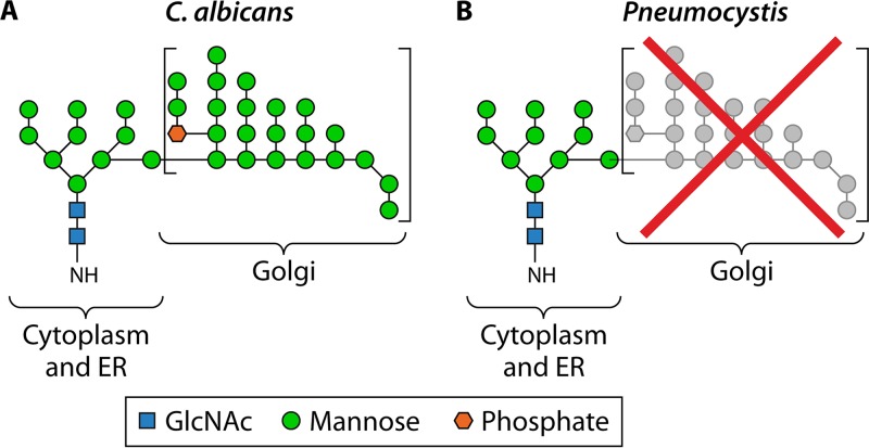
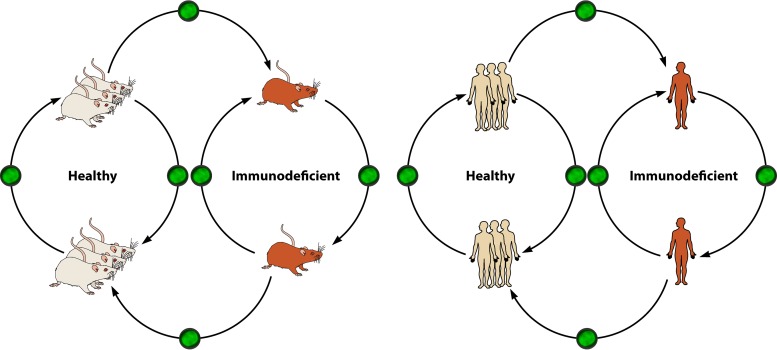
Pneumocystis, a unique atypical fungus with an elusive lifestyle, has had an important medical history. It came to prominence as an opportunistic pathogen that not only can cause life-threatening pneumonia in patients with HIV infection and other immunodeficiencies but also can colonize the lungs of healthy individuals from a very early age. The genus Pneumocystis includes a group of closely related but heterogeneous organisms that have a worldwide distribution, have been detected in multiple mammalian species, are highly host species specific, inhabit the lungs almost exclusively, and have never convincingly been cultured in vitro, making Pneumocystis a fascinating but difficult-to-study organism. Improved molecular biologic methodologies have opened a new window into the biology and epidemiology of Pneumocystis. Advances include an improved taxonomic classification, identification of an extremely reduced genome and concomitant inability to metabolize and grow independent of the host lungs, insights into its transmission mode, recognition of its widespread colonization in both immunocompetent and immunodeficient hosts, and utilization of strain variation to study drug resistance, epidemiology, and outbreaks of infection among transplant patients. This review summarizes these advances and also identifies some major questions and challenges that need to be addressed to better understand Pneumocystis biology and its relevance to clinical care.

Keywords: Pneumocystis; epidemiology; genome features; molecular biology; strain variation; transmission.


Figures

FIG 1
FIG 1
Phylogenetic relationships and ortholog conservation for Pneumocystis and related fungi. The maximum likelihood tree was inferred from 248 single-copy core orthologs. Ortholog conservation patterns highlighted include core orthologs found in all genomes (CORE), ascomycete-specific orthologs found in all ascomycetes but not in Microsporidia (ASCOM), basidiomycete-specific orthologs found in both Cryptococcus neoformans and Ustilago maydis but not in all other fungi (BASIDIO), Pneumocystis-specific orthologs (PNEUMO), orthologs shared in any two or more genomes (SHARED), and orthologs unique to only one genome (UNIQUE). Numbers on the branches of the tree indicate bootstrap support values. The phyla and subphyla are indicated on the main branches as follows: Ta, Taphrinomycotina; Sa, Saccharomycotina; Pe, Pezizomycotina; Ba, Basidiomycota; and Mi, Microsporidia.
FIG 2
FIG 2
Top enriched and depleted protein families in Pneumocystis. Significantly enriched and depleted Pfam domains (Fisher's exact test; q < 0.05) were included in the heat map if the domains appeared at least twice in the following comparisons: Pneumocystis versus Schizosaccharomyces, Pneumocystis versus Schizosaccharomyces and Taphrina deformans, Pneumocystis versus S. cerevisiae and C. albicans, Pneumocystis versus Encephalitozoon cuniculi and E. intestinalis, and Pneumocystis versus all others shown. CFEM, common in fungal extracellular membrane domain; RRM, RNA recognition motif. The heat map is color coded based on Z scores from −2 to 3, as indicated by the key. Fungal species are ordered based on their phylogenetic relationships, as indicated at the bottom. The subphyla are indicated on the main branches, as follows: TA, Taphrinomycotina; SA, Saccharomycotina; and MI, Microsporidia. (Modified from Fig. 2 in reference .)
FIG 3
FIG 3
Cell wall structure of Pneumocystis compared to that of C. albicans. (A) C. albicans cell wall. The inner layer contains chitin and β-glucans, whereas the outer layer contains hypermannosylated N- and O-linked glycans (mannans) that are covalently linked with proteins to form glycoproteins. The plasma membrane contains ergosterol. (Electron micrograph courtesy of Louise Walker and Neil Gow, University of Aberdeen, United Kingdom; reprinted with permission.) (B) Cell wall of Pneumocystis cysts (asci). The inner layer contains β-glucans and no chitin, whereas the outer layer is highly enriched in proteins that are glycosylated via N- and O-linked glycans, but without the mannan outer chains. The plasma membrane contains cholesterol instead of ergosterol. (C) Cell wall of Pneumocystis trophic forms. The cell wall is the same as that of Pneumocystis cysts, except for the absence of β-glucans.
FIG 4
FIG 4
Loss of chitin in Pneumocystis cell wall. P. murina-infected lung tissue (A) and C. albicans-infected kidney tissue (positive control) (C) were stained with a recombinant chitin binding domain (green). Chitin staining is absent in P. murina (A) but readily detected in C. albicans (C). (B) Pneumocystis organisms are demonstrated by dual staining with anti-Msg (red), which labels both trophic forms and cysts, and dectin-Fc (green), which labels β-1,3-glucan in cysts. Original magnification, ×400.
FIG 5
FIG 5
Lack of N-linked hypermannose (mannan) outer chains in Pneumocystis. Like C. albicans, Pneumocystis spp. are able to synthesize the N-linked glycan core structure (containing up to nine mannose residues, as indicated on the left) in the cytoplasm and the endoplasmic reticulum (ER). However, due to the loss of multiple enzymes, Pneumocystis spp. are unable to synthesize the α-1,6-linked mannose backbone as well as the α-1,2- and α-1,3-linked mannose outer chains seen in C. albicans (square brackets), which are synthesized in the Golgi apparatus. (Diagrams of the N-linked mannan structure in C. albicans and Pneumocystis were adapted from reference .)
FIG 6
FIG 6
High intron densities in Pneumocystis genomes. The graphs show numbers of introns per gene as a function of gene length (measured in kilobases) for three Pneumocystis species as well as for the fission yeast Schizosaccharomyces pombe. Each dot represents a single gene. Intron densities per gene for 1,624 orthologous genes are systematically higher for Pneumocystis spp. than those for S. pombe. Intron positions and sizes were extracted from annotated GenBank files for P. jirovecii (accession no. GCA_001477535.1) in panel A, P. carinii (accession no. GCA_001477545.1) in panel B, P. murina (accession no. GCF_000349005.1) in panel C, and S. pombe (accession no. GCF_000002945.1) in panel D.
FIG 7
FIG 7
msg-RFLP analysis of P. jirovecii. Msg is encoded by a multicopy msg gene family (msg-A1 subfamily) in P. jirovecii, with an estimated 80 to 90 variable copies per genome. DNAs were extracted from respiratory samples from patients with PCP. The downstream region of the msg gene repertoire was amplified by PCR, using primers targeting conserved regions, followed by restriction digestion with the enzyme DraI and then electrophoresis in conventional agarose gels stained with SYBR green. Labels at the top represent the DNA marker (lane M) and individual patient samples (numbered lanes). (A) Samples from different HIV-infected patients with PCP, except for samples 4a and 4b, which were sequential samples from the same patient. (B) Samples from different renal transplant patients with PCP from an outbreak in Germany (345). Note that the RFLP patterns among samples from unrelated HIV patients are different from each other, whereas the RFLP patterns among the renal transplant patients are identical to each other.
FIG 8
FIG 8
Quantification of the tandem repeat copy number in the msg-UCS of P. jirovecii. The tandem repeat region in the intron of the single-copy msg-UCS gene was amplified by PCR, separated in an acrylamide sizing gel, and stained by silver staining as described by Ma et al. (300). Numbers above each lane represent individual patients. Lanes M contain a DNA size marker, with the number of repeats indicated above each DNA band. Each band within a lane represents a unique Pneumocystis strain identified in that patient. For example, lane 1 was obtained from a patient infected with three strains, lane 2 from a patient infected with two strains, and lane 5 from a patient infected with a single strain.
FIG 9
FIG 9
Hypothetical transmission mode of Pneumocystis in animals and humans. In this mode, cysts (indicated by green, as seen with immunofluorescence staining) serve as the infectious form and mammalian hosts (e.g., rodents on the left and humans on the right) as the reservoir for infection; transmission occurs via the airborne route between immunocompetent hosts, between immunodeficient hosts, or between immunocompetent and immunodeficient hosts. The healthy population (indicated by three individuals) is substantially larger than the immunodeficient population (indicated by one individual).
None
None
None

References

    1. Beard CB, Fox MR, Lawrence GG, Guarner J, Hanzlick RL, Huang L, del Rio C, Rimland D, Duchin JS, Colley DG. 2005. Genetic differences in Pneumocystis isolates recovered from immunocompetent infants and from adults with AIDS: epidemiological implications. J Infect Dis 192:1815–1818. doi:10.1086/497381. - DOI - PubMed
    1. Vargas SL, Hughes WT, Santolaya ME, Ulloa AV, Ponce CA, Cabrera CE, Cumsille F, Gigliotti F. 2001. Search for primary infection by Pneumocystis carinii in a cohort of normal, healthy infants. Clin Infect Dis 32:855–861. doi:10.1086/319340. - DOI - PubMed
    1. Gajdusek DC. 1957. Pneumocystis carinii; etiologic agent of interstitial plasma cell pneumonia of premature and young infants. Pediatrics 19:543–565. - PubMed
    1. Goldman AS, Goldman LR, Goldman DA. 2005. What caused the epidemic of Pneumocystis pneumonia in European premature infants in the mid-20th century? Pediatrics 115:e725–e736. doi:10.1542/peds.2004-2157. - DOI - PubMed
    1. Centers for Disease Control and Prevention. 1982. A cluster of Kaposi's sarcoma and Pneumocystis carinii pneumonia among homosexual male residents of Los Angeles and Orange Counties, California. MMWR Morb Mortal Wkly Rep 31:305–307. - PubMed

Publication types

MeSH terms

LinkOut - more resources