. 2018 Jun 13;10(445):eaao2586.
doi: 10.1126/scitranslmed.aao2586.

Probiotic strains detect and suppress cholera in mice

Affiliations

Probiotic strains detect and suppress cholera in mice

Ning Mao et al. Sci Transl Med. .

Abstract

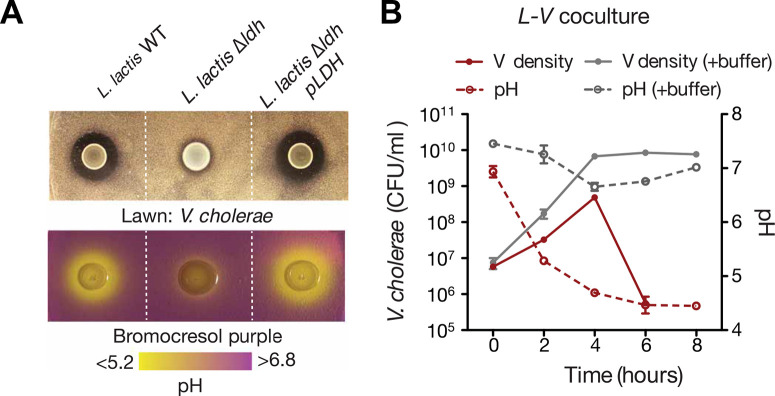
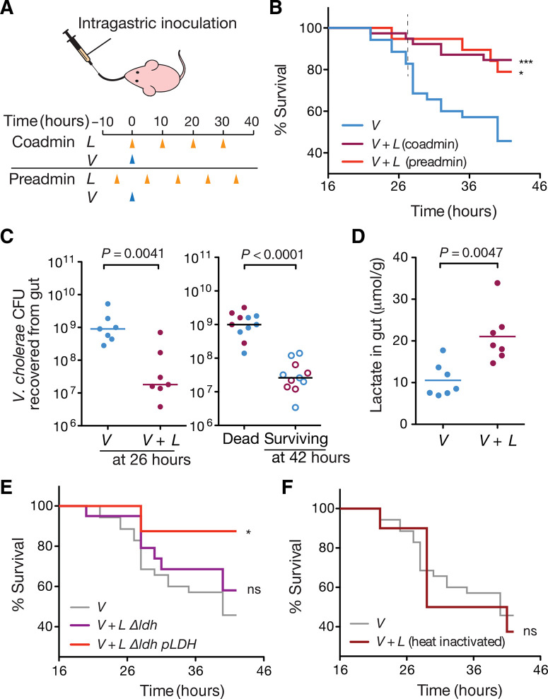
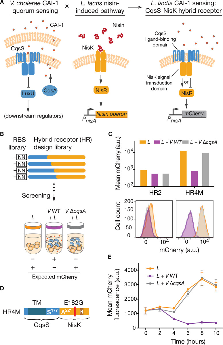
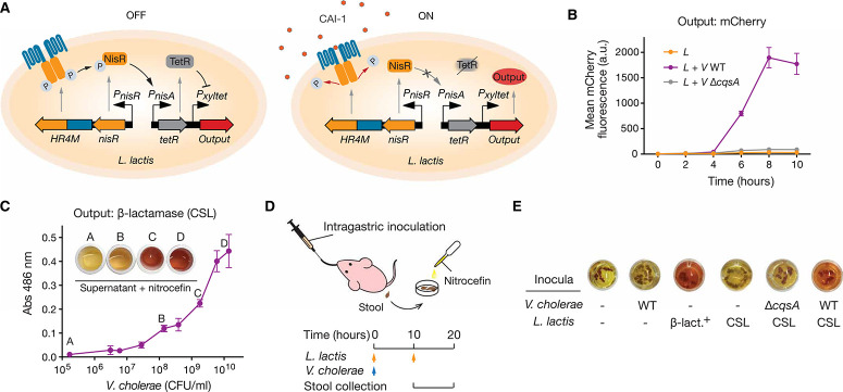
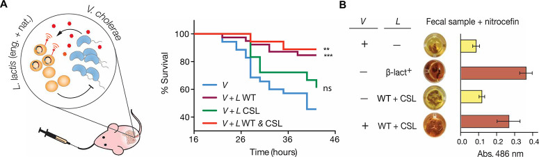
Microbiota-modulating interventions are an emerging strategy to promote gastrointestinal homeostasis. Yet, their use in the detection, prevention, and treatment of acute infections remains underexplored. We report the basis of a probiotic-based strategy to promote colonization resistance and point-of-need diagnosis of cholera, an acute diarrheal disease caused by the pathogen Vibrio cholerae Oral administration of Lactococcus lactis, a common dietary fermentative bacterium, reduced intestinal V. cholerae burden and improved survival in infected infant mice through the production of lactic acid. Furthermore, we engineered an L. lactis strain that specifically detects quorum-sensing signals of V. cholerae in the gut and triggers expression of an enzymatic reporter that is readily detected in fecal samples. We postulate that preventive dietary interventions with fermented foods containing natural and engineered L. lactis strains may hinder cholera progression and improve disease surveillance in populations at risk of cholera outbreaks.


Figures

Fig. 1
Fig. 1
Lactic acid–dependent antibacterial effect of L. lactis against V. cholera in vitro. (A) Top: Agar diffusion assay of wild-type (WT), lactate dehydrogenase mutant (Δldh), and plasmid-complemented lactate dehydrogenase mutant Δldh (pLDH) strains of L. lactis grown on a lawn of V. cholerae. Bottom: Agar diffusion assay of L. lactis strains in minimally buffered GM17 agar plates containing pH indicator bromocresol purple, which turns yellow when pH drops below 5.2. Inhibition zones correlate with the acidification range of L. lactis colonies. (B) Acidification dynamics and V. cholerae cell density in coculture with L. lactis in minimally buffered media and strongly buffered media (+buffer). For detailed media conditions, see Materials and Methods and fig. S2. Bars represent range of technical duplicates.
Fig. 2
Fig. 2
L. lactis–derived lactic acid antagonizes V. cholerae infection in mice. (A) Intragastric feeding regimens of L. lactis and inoculation time of V. cholerae to infant mice. Mock-fed mice were administered GM17 medium instead of L. lactis. (B) Effects of L. lactis intervention on infant mouse survival to cholera infection under both feeding regimens. Coadministration with V. cholerae, ***P = 0.0005; preadministration, *P = 0.0187, log-rank test against mock-fed (V). V: n = 37, N = 6; V + L, coadmin: n = 39, N = 6; V + L, preadmin: n = 19, N = 6. (C) Effect of L. lactis intervention on V. cholerae colonization in the infant mouse gut. (C) Left: V. cholerae intestinal colonization at 26 hours after infection. Right: V. cholerae burden in mice that died from cholera and in mice that survived the infection by 42 hours after infection. Each data point represents an individual mouse. Horizontal lines are medians. P values from Mann-Whitney test (n = 7, N = 7 for nontreated/treated; n = 11, N = 7 for dead versus survived, respectively). Data correspond to coadministration regimen. (D) Intestinal lactate concentration in L. lactis–treated and mock-treated infant mice at 26 hours after infection. Mann-Whitney test (n = 7, N = 7). Data correspond to coadministration regimen. (E) Effects of lactate dehydrogenase mutant (Δldh) and plasmid-complemented lactate dehydrogenase (Δldh pLDH) L. lactis strains on infant mouse survival to cholera infection. Functional knockout of lactate dehydrogenase compromises the protective effect against cholera (n = 20, N = 6). ns, not significant by log-rank test compared to mock treatment (V). Complementation with a plasmid-based ldh restores the protective effect (*P = 0.0349, log-rank test; n = 9, N = 3). (F) Effect of treatment with heat-inactivated L. lactis on infant mice survival to cholera infection. n = 10, N = 3. n, number of individual mice; N, number of litters covered in each group.
Fig. 3
Fig. 3
Construction of HR for V. cholerae sensing. (A) Creation of a CAI-1–sensing function in L. lactis requires the fusion of two distantly related quorum-sensing signaling pathways. The proposed HR fuses the CAI-1–binding domain of the CqsS receptor with the histidine kinase domain of NisK to achieve CAI-1–dependent signaling in L. lactis. The design of the HR cannot predict whether the regulatory outcome of CAI-1 binding will be activation (arrow-headed line) or inactivation of NisR (bar-headed line). (B) Functional screen for HR variants. CqsS-NisK fusion variants with randomized RBS strengths (fig. S9) were screened for their ability to modulate output gene expression in response to CAI-1. CAI-1–deficient V. cholerae (V ΔcqsA) serves as a negative control. (C) mCherry fluorescence output of HR variants in response to CAI-1. Hybrid receptor 4 mutant (HR4M) is a functional variant. Hybrid receptor 2 (HR2) is an example of a nonfunctional variant. (D) Primary sequence map of HR4M. S177 indicates the last residue of N-terminal part of CqsS, and A221 indicates the first residue of the C-terminal part of NisK. The HR contains a spontaneous Glu-to-Gly mutation at residue 182. (E) Reporter gene expression dynamics in L. lactis with HR4M in response to CAI-1. Error bars are SEM of three technical replicates. a.u., arbitrary units.
Fig. 4
Fig. 4
Cholera sensing and reporting by engineered L. lactis in vitro and in vivo. (A) Engineered CAI-1–dependent signaling in L. lactis. The HR4M-NisR two-component system sensing module and a TetR/Pxyltet signal-processing module constitute the complete V. cholerae–sensing circuit. In the absence of CAI-1, constitutive expression of TetR prevents the expression of the output gene. In the presence of CAI-1, the hybrid two-component system stops its phosphorelay, halting TetR expression and leading to activation of the output gene. (B) Activation dynamics of the cholera-sensing circuit. Error bars are SEM of three technical replicates. (C) Colorimetric reporting of V. cholerae sensing. The L. lactis CSL strain secretes β-lactamase, which hydrolyzes nitrocefin and produces a visible color change from yellow to red (maximum absorbance at 486 nm). Bars represent range of absorbance for technical duplicates. (D) Schematic of the infant mouse model for in vivo testing of the cholera-sensing strain. Cholera-infected mice were fed with L. lactis CSL, and stools were collected between 10 and 20 hours after infection for nitrocefin-based β-lactamase assay. (E) Performance of the living diagnostic and β-lactamase assay for the detection of cholera. Color change in nitrocefin with fecal samples indicates β-lactamase activity. V. cholerae WT, CAI-1–deficient V. cholerae (V ΔcqsA), and L. lactis cholera-sensing Lactococcus (CSL) strain. Constitutive β-lactamase expression L. lactis (β-lact.+).
Fig. 5
Fig. 5
L. lactis intervention for combined cholera protection and detection. (A) Left: Strategy of mixed population of natural and engineered L. lactis for cholera protection and detection. The natural lactic acid bacteria acidify the intestinal environment to antagonize the proliferation of the pathogen. The engineered probiotics detect the quorum-sensing molecule of V. cholerae and produce an easy-to-read reporter. Right: Infant mouse survival to V. cholerae infection after the intervention with single or combined L. lactis strains. n = 18, N = 4 for CSL and WT + CSL, respectively. V and V + L WT curves are overlays from Fig. 2B. Log-rank test against the mock-fed group (V): ***P = 0.005, WT coadministration; **P = 0.0034, WT and CSL; ns, CSL. (B) In vivo detection of V. cholerae using single or combined doses of L. lactis strains. Dosing scheme was performed as described in Fig. 4D. Absorbance at 486 nm was measured on three independent samples in each group. Images on the left column correspond to one representative sample. Error bars represent SEM.

Comment in

  • Curbing cholera.
    Hall RH. Hall RH. Sci Transl Med. 2018 Jun 13;10(445):eaat9483. doi: 10.1126/scitranslmed.aat9483. Sci Transl Med. 2018. PMID: 29899025

References

    1. Ali M, Nelson A. R, Lopez A. L, Sack D. A, Updated global burden of cholera in endemic countries. PLOS Negl. Trop. Dis. 9, e0003832 (2015). - PMC - PubMed
    1. Marteau P. R, de Vrese M, Cellier C. J, Schrezenmeir J, Protection from gastrointestinal diseases with the use of probiotics. Am. J. Clin. Nutr. 73, 430S–436S (2001). - PubMed
    1. Kamada N, Chen G. Y, Inohara N, Núñez G, Control of pathogens and pathobionts by the gut microbiota. Nat. Immunol. 14, 685–690 (2013). - PMC - PubMed
    1. Hsiao A, Ahmed A. M. S, Subramanian S, Griffin N. W, Drewry L. L, Petri W. A Jr., Haque R, Ahmed T, Gordon J. I, Members of the human gut microbiota involved in recovery from Vibrio cholerae infection. Nature 515, 423–426 (2014). - PMC - PubMed
    1. Duan F., March J. C, Engineered bacterial communication prevents Vibrio cholera virulence in an infant mouse model. Proc. Natl. Acad. Sci. U.S.A. 107, 11260–11264 (2010). - PMC - PubMed

Publication types

LinkOut - more resources