. 2018 Jul 31;92(16):e00838-18.
doi: 10.1128/JVI.00838-18. Print 2018 Aug 15.

Multiple Host Factors Interact with the Hypervariable Domain of Chikungunya Virus nsP3 and Determine Viral Replication in Cell-Specific Mode

Affiliations

Multiple Host Factors Interact with the Hypervariable Domain of Chikungunya Virus nsP3 and Determine Viral Replication in Cell-Specific Mode

Chetan D Meshram et al. J Virol. .

Abstract

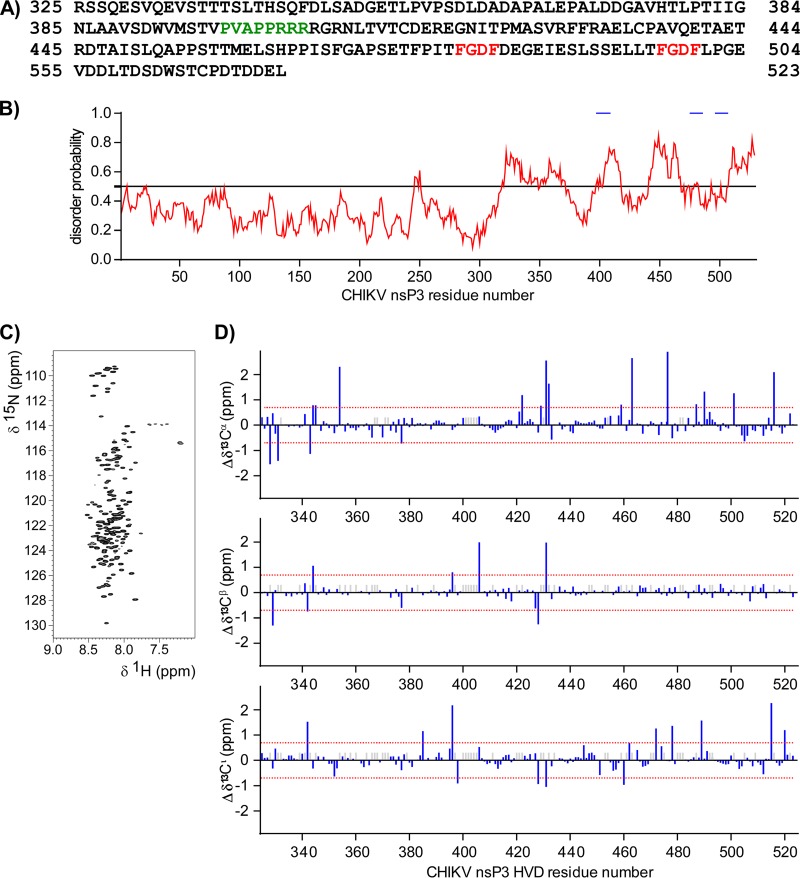
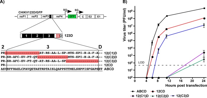
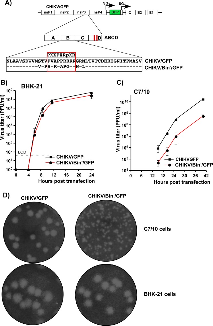
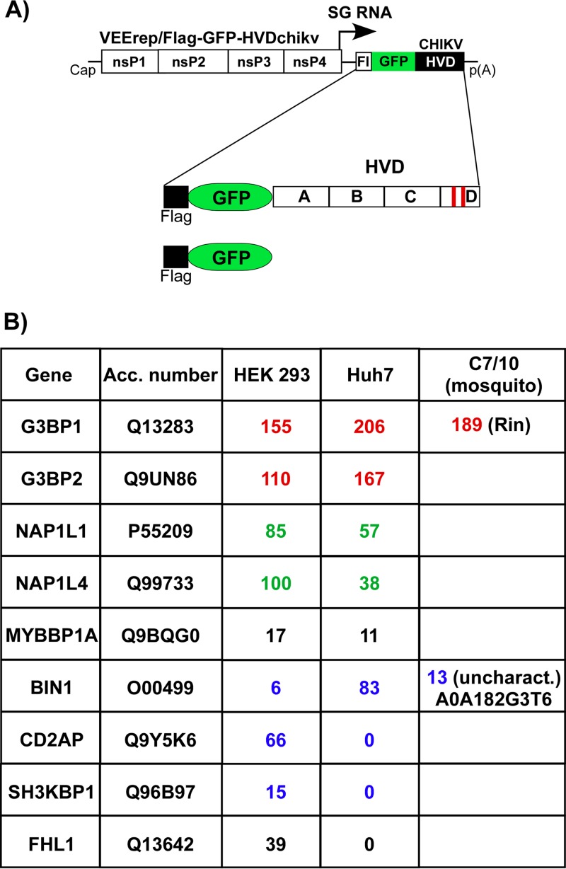
Alphaviruses are widely distributed in both hemispheres and circulate between mosquitoes and amplifying vertebrate hosts. Geographically separated alphaviruses have adapted to replication in particular organisms. The accumulating data suggest that this adaptation is determined not only by changes in their glycoproteins but also by the amino acid sequence of the hypervariable domain (HVD) of the alphavirus nsP3 protein. We performed a detailed investigation of chikungunya virus (CHIKV) nsP3 HVD interactions with host factors and their roles in viral replication in vertebrate and mosquito cells. The results demonstrate that CHIKV HVD is intrinsically disordered and binds several distinctive cellular proteins. These host factors include two members of the G3BP family and their mosquito homolog Rin, two members of the NAP1 family, and several SH3 domain-containing proteins. Interaction with G3BP proteins or Rin is an absolute requirement for CHIKV replication, although it is insufficient to solely drive it in either vertebrate or mosquito cells. To achieve a detectable level of virus replication, HVD needs to bind members of at least one more protein family in addition to G3BPs. Interaction with NAP1L1 and NAP1L4 plays a more proviral role in vertebrate cells, while binding of SH3 domain-containing proteins to a proline-rich fragment of HVD is more critical for virus replication in the cells of mosquito origin. Modifications of binding sites in CHIKV HVD allow manipulation of the cell specificity of CHIKV replication. Similar changes may be introduced into HVDs of other alphaviruses to alter their replication in particular cells or tissues.IMPORTANCE Alphaviruses utilize a broad spectrum of cellular factors for efficient formation and function of replication complexes (RCs). Our data demonstrate for the first time that the hypervariable domain (HVD) of chikungunya virus nonstructural protein 3 (nsP3) is intrinsically disordered. It binds at least 3 families of cellular proteins, which play an indispensable role in viral RNA replication. The proteins of each family demonstrate functional redundancy. We provide a detailed map of the binding sites on CHIKV nsP3 HVD and show that mutations in these sites or the replacement of CHIKV HVD by heterologous HVD change cell specificity of viral replication. Such manipulations with alphavirus HVDs open an opportunity for development of new irreversibly attenuated vaccine candidates. To date, the disordered protein fragments have been identified in the nonstructural proteins of many other viruses. They may also interact with a variety of cellular factors that determine critical aspects of virus-host interactions.

Keywords: BIN1; CD2AP; Eilat virus; G3BP1; G3BP2; NAP1L1; alphavirus; chikungunya virus; viral RNA replication; virus-host interactions.


Figures

FIG 1
FIG 1
CHIKV HVD is an intrinsically disordered domain. (A) Amino acid sequence of CHIKV 181/25 nsP3 HVD. The binding motif for BIN1 is depicted in green. The binding motifs for G3BP1 and G3BP2 are depicted in red. (B) Computational prediction of CHIKV nsP3 disorder probability by IUPred. The known binding motifs for BIN1 and G3BPs are marked by blue lines. (C) 2D best-TROSY spectra of CHIKV nsP3 HVD showing cross peaks corresponding to the one-bond interaction between proton, 1H, and nitrogen, 15N, of the amide bonds. (D) Results of chemical shift analysis of 13C′, 13Cα, and 13Cβ nuclei in CHIKV HVD. The gray bars indicate unassigned carbons.
FIG 2
FIG 2
Deletions of G3BP-binding peptides in CHIKV nsP3 HVD have deleterious effects on viral replication. (A) Schematic presentation of CHIKV/GFP genome, sequences of the carboxy-terminal fragments of nsP3 HVD in the parental CHIKV/GFP and in the designed deletion mutants, and infectivities of the in vitro-synthesized RNAs in ICA performed on BHK-21 and C7/10 cells. The repeating element is indicated in red. Dots indicate deleted aa in the designed mutants. NV, not viable. The numbers given are the aa numbers in the original full-length CHIKV nsP3. (B and C) BHK-21 and C7/10 cells, respectively, were electroporated with 3 μg of the indicated in vitro-synthesized RNAs, and samples of the media were collected at the indicated time points. Virus titers were determined by plaque assay on BHK-21 cells. The experiments were performed twice using different amounts of in vitro-synthesized RNA and demonstrated similar differences in the rates of infectious virus release. The results of one of the experiments are presented.
FIG 3
FIG 3
CHIKV nsP3 HVD fragments other than the carboxy-terminal, G3BP-binding repeat determine virus replication. (A) Schematic presentation of the parental CHIKV/GFP genome and its derivative, CHIKV/artHVD/GFP, with artificially designed HVD, and alignment of the wt and mutated HVD sequences. The repeating element is indicated in red. Dashes indicate identical aa. (B) Computational prediction of disorder probability of CHIKV nsP3 with artHVD (shown in blue) and wt CHIKV nsP3 (shown in red) by IUPred. The mutated HVD fragment is indicated by a green line. (C) Replication rates of the indicated viruses, CHIKV/GFP, CHIKV/artHVD/GFP, and its derivative CHIKV/artHVD(K258I)/GFP, which contained an adaptive mutation, K258I, in nsP3. BHK-21 cells were electroporated with 3 μg in vitro-synthesized RNAs. Viral samples were harvested at the indicated time points, and titers were determined by plaque assay on BHK-21 cells. LOD, limit of detection. (D) Titers of CHIKV/GFP and CHIKV/artHVD(K258I)/GFP at 24 h after infection of the indicated cell lines at an MOI of 1 PFU/cell. Titers were determined by plaque assay on BHK-21 cells. The results of one of the reproducible experiments performed in triplicates are presented. Means and standard deviations (SDs) are indicated.
FIG 4
FIG 4
Fragments of CHIKV nsP3 HVD differentially regulate viral replication. (A) Alignment of amino acid sequences of wt ABCD HVD and mutated 123D HVD counterpart. Fragments A, B, and C in wt HVD and the corresponding mutated fragments 1, 2, and 3 are indicated by black boxes. Red boxes indicate positions of the G3BP-binding sites. Dashes indicate identical aa. (B) Schematic presentation of the original CHIKV/GFP genome and modified HVDs in the genomes of the designed viruses. Equal amounts (3 μg) of the in vitro-synthesized RNAs of the indicated constructs were electroporated into BHK-21 cells, and RNA infectivities and plaque sizes were compared in the ICA. (C) Schematic presentation of HVDs of the same viruses as in panel B, shown for clarity of presentation. Equal amounts of the in vitro-synthesized RNAs of the indicated constructs were electroporated into C7/10 cells, and RNA infectivities and plaque sizes were compared in the ICA performed on C7/10 cells. NV, not viable. (D) Replication rates of the indicated variants were determined in the experiment presented in panel B. Samples were harvested at the indicated time points, and viral titers were determined by plaque assay on BHK-21 cells. The experiments were repeated multiple times using different sets of RNAs with consistent results. (E) Replication rates of the indicated variants were determined in the experiment presented in panel C. At the indicated time points, viral titers were determined by plaque assay on C7/10 cells. The experiments were repeated multiple times using different sets of RNAs with consistent results. The experiments presented in panels D and E show the data of the most representative experiments that used all the indicated variants together. (F) Titers of multiple, randomly selected samples of the indicated viruses harvested from BHK-21 and C7/10 cells were determined in parallel on both BHK-21 and C7/10 cells. The ratios of the determined titers are presented as means and SDs.
FIG 5
FIG 5
The carboxy terminus of fragment C determines CHIKV replication in vertebrate cells. (A) Schematic presentation of CHIKV/123D/GFP genome and aa sequences of fragment 3 that contained the peptides of mutated HVD restored to wt sequence. The restored fragments are indicated in red, and dashes indicate aa that are identical to those in wt CHIKV nsP3 HVD. (B) BHK-21 cells were electroporated with 3 μg of the in vitro-synthesized RNAs of the indicated constructs. Media were replaced at the indicated time points, and viral titers were determined by plaque assay on BHK-21 cells. The experiments were repeated three times with highly reproducible results. LOD, limit of detection.
FIG 6
FIG 6
Proline-rich, SH3-binding motif in CHIKV HVD is more important for viral replication in mosquito than in vertebrate cells. (A) Schematic presentation of CHIKV/GFP genome and its derivative CHIKV/Bin/GFP with mutated SH3-binding motif. Dashes in the alignment indicate aa that are identical in wt and mutated CHIKV nsP3 HVD. (B and C) BHK-21 and C7/10 cells, respectively, were electroporated with 3 μg of in vitro-synthesized RNAs. At the indicated time points, media were replaced and viral titers were determined by plaque assay on BHK-21 cells. LOD, limit of detection. These experiments were repeated twice using different amounts of RNA and produced consistent results. (D) Size of plaques formed by parental CHIKV/GFP and its CHIKV/Bin/GFP derivative on BHK-21 and C7/10 cells. For both cell lines, plaques were stained by crystal violet at 3 days postinfection.
FIG 7
FIG 7
Host factors binding to CHIKV HVD in different cell types. (A) Schematic presentation of VEEV replicons that encode Flag-GFP-HVDchikv fusion or Flag-GFP. (B) Indicated cell lines were infected with packaged VEErep/Flag-GFP-HVDchikv replicon or the control replicon expressing only Flag-GFP at an MOI of 20 IU/cell. Cells were harvested at 2 h postinfection, and complexes were immunoprecipitated as described in Materials and Methods. Proteins were separated by SDS-PAGE and identified by mass spectrometry. Total spectra for the most abundant proteins are presented. Groups of functionally similar proteins are indicated by different colors. None of the presented proteins were detected in the control samples generated using Flag-GFP. Acc. number, accession number for UniProt database.
FIG 8
FIG 8
Mapping of G3BP-, NAP1-, and CD2AP-binding sites in CHIKV nsP3 HVD. (A) Schematic presentation of VEEV replicon and expressed Flag-GFP-HVD cassettes that contained wt and mutated CHIKV HVD fragments. Fragments with wt aa sequences are indicated by open boxes and labeled A, B, C, and D, and mutated counterparts of A, B, and C are indicated by black boxes and labeled 1, 2, and 3. (B, C, D, and E) Indicated cell lines were infected with packaged replicons encoding indicated fragments of CHIKV HVD. At 2 h postinfection, protein complexes were isolated using magnetic beads with Flag-specific MAbs as described in Materials and Methods. The presence of studied cellular proteins was analyzed by Western blotting using specific Abs and corresponding secondary Abs labeled with different infrared dyes. Membranes were scanned on a Li-Cor imager. The following primary antibodies were used: anti-G3BP2 rabbit polyclonal antibodies (A302-040; Epitomics), anti-CD2AP rabbit polyclonal antibodies (sc-9137; Santa Cruz), anti-NAP1L1 rabbit polyclonal antibodies (14989-1-AP; Proteintech), and anti-Flag mouse monoclonal antibodies (F1804; Sigma).
FIG 9
FIG 9
CHIKV and EILV HVDs determine cell specificity of viral replication. (A) Alignment of the aa sequences of CHIKV and EILV HVDs. Dots indicate gaps introduced for better alignment. Dashes indicate identical aa. Red boxes indicate positions of the carboxy-terminal, G3BP/Rin-binding repeats. (B) Schematic presentations of recombinant alphavirus CHIKV and EILV/CHIKV genomes with homologous and heterologous nsP3 HVDs. The white and black boxes indicate CHIKV and EILV genomes, respectively. Titers of rescued viruses after electroporation of 3 μg of the in vitro-synthesized RNAs into indicated cell lines. Titers of the stocks were determined by plaque assay on both BHK-21 and C7/10 cells. NV, not viable. Titers of plaque-forming viruses are shown as PFU/ml, and titers of noncytopathic viruses were measured in GFP-positive focus-forming units (FFU/ml). (C) BHK-21 and C7/10 cells were infected with the indicated viruses, which were rescued by electroporation of permissive cells. Images were taken on a fluorescence microscope.
FIG 10
FIG 10
Addition of CHIKV HVD fragment C to EILV HVD makes the resulting chimeric CHIKV/HVDeilv(C)/GFP capable of replication in vertebrate cells. (A) Schematic presentation of recombinant CHIKV genomes with wt or heterologous HVDs, infectivity of the in vitro-synthesized RNA in ICA on BHK-21 cells, and titers of viruses harvested 24 h after electroporation of BHK-21 cells. (B) Replication rates of the indicated viruses in BHK-21 cells after electroporation of the in vitro-synthesized RNAs. Viral titers at the indicated time points were determined by plaque assay on BHK-21 cells. (C) GFP expression by replicating viruses in BHK-21 and C7/10 cells. Cells were infected with the indicated viruses at an MOI of 5 PFU/cell. Stocks of CHIKV/GFP and CHIKV/HVDeilv(C)/GFP used for infection were generated on BHK-21 cells, and the stock of CHIKV/HVDeilv/GFP was generated on C7/10 cells. Images were acquired on a fluorescence microscope at 8 h and 16 h after infection of BHK-21 and C7/10 cells, respectively.
FIG 11
FIG 11
Locations of to-date identified binding sites of cellular proteins in CHIKV nsP3 HVD. CHIKV nsP3 is structurally divided into three domains, which include macro domain, AUD, and HVD. In this study, HVD was further divided into the indicated four fragments, A, B, C, and D. The identified locations of binding sites for cellular proteins are indicated in different colors. A question mark indicates that a possibility remains that CD2AP requires interaction with more than one HVD motif for efficient binding. “3” indicates a position of a noncanonical G3BP-binding site in C3 peptide of CHIKV HVD.

Similar articles

Cited by

References

    1. Griffin DE. 2001. Alphaviruses, p 917–962. In Knipe DM, Howley PM, Griffin DE, Lamb RA, Martin MA, Roizman B, Straus SE (ed), Fields virology, 4th ed Lippincott Williams & Wilkins, Philadelphia, PA.
    1. Strauss JH, Strauss EG. 1994. The alphaviruses: gene expression, replication, evolution. Microbiol Rev 58:491–562. - PMC - PubMed
    1. Weaver SC, Frolov I. 2005. Togaviruses, p 1010–1024. In Mahy BWJ, ter Meulen V (ed), Virology, vol 2 Hodder Arnold, Salisbury, United Kingdom.
    1. Weaver SC, Lecuit M. 2015. Chikungunya virus infections. N Engl J Med 373:94–95. doi:10.1056/NEJMc1505501. - DOI - PubMed
    1. Weaver SC, Lecuit M. 2015. Chikungunya virus and the global spread of a mosquito-borne disease. N Engl J Med 372:1231–1239. doi:10.1056/NEJMra1406035. - DOI - PubMed

Publication types

MeSH terms

Substances