Review
. 2018 Jun 13;16(6):208.
doi: 10.3390/md16060208.

Neuronal Nicotinic Acetylcholine Receptor Modulators from Cone Snails

Affiliations
Review

Neuronal Nicotinic Acetylcholine Receptor Modulators from Cone Snails

Nikita Abraham et al. Mar Drugs. .

Abstract

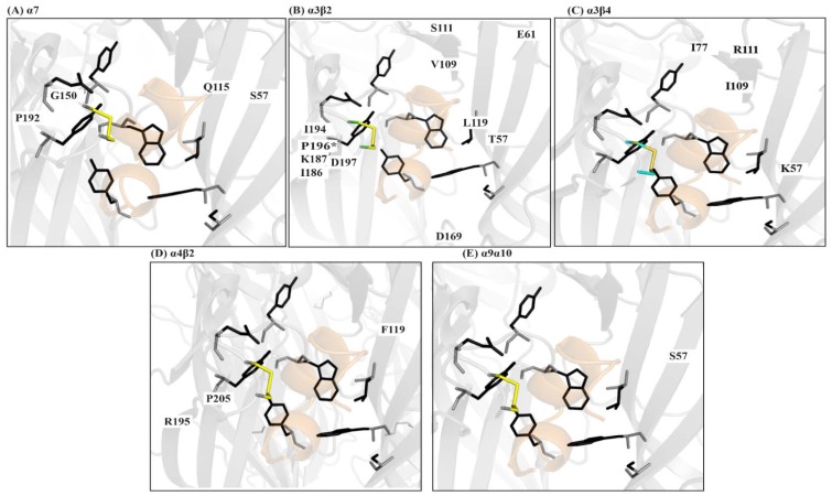
Marine cone snails are a large family of gastropods that have evolved highly potent venoms for predation and defense. The cone snail venom has exceptional molecular diversity in neuropharmacologically active compounds, targeting a range of receptors, ion channels, and transporters. These conotoxins have helped to dissect the structure and function of many of these therapeutically significant targets in the central and peripheral nervous systems, as well as unravelling the complex cellular mechanisms modulated by these receptors and ion channels. This review provides an overview of α-conotoxins targeting neuronal nicotinic acetylcholine receptors. The structure and activity of both classical and non-classical α-conotoxins are discussed, along with their contributions towards understanding nicotinic acetylcholine receptor (nAChR) structure and function.

Keywords: conotoxins; nicotinic acetylcholine receptors; α-conotoxins.


Conflict of interest statement

The authors declare no conflict of interest

Figures

Figure 1
Figure 1
Conotoxin expression. (A) Conotoxins are produced in the venom apparatus that consists of (1) the venom bulb—a muscular organ that is used as a pump, (2) the venom duct which produces the venom, (3) the radular sac that stores the harpoons. Harpoons are evolutionarily modified radula teeth that are used to inject venom into the prey, like a hypodermic needle, (4) the proboscis is used to “load” (5) the harpoon at the tip to inject venom; and, (B) The venom peptides are translated as prepropeptide. They consist of a conserved N-terminal made up of the signal sequence and propeptide region which are cleaved during peptide processing. The final bioactive conotoxin consists of only the C-terminal region, which is also the disulfide rich hypervariable region of the prepropeptide.
Figure 2
Figure 2
Conotoxin structural and functional diversity. (A) Conotoxins have highly diverse sequences, cysteine frameworks, connectivity, and biological targets. χ-MrIA and α-TxIA have the same cysteine framework, but different connectivities and sequence which contribute to the difference in their targets. On the contrary, ω-MVIIC and μO-MrVIB have the same cysteine framework and connectivity but different biological targets. (O = hydroxyproline); (B) Conotoxin diversity extends to their three-dimensional structures, and thereby further adding to their functional diversity.
Figure 3
Figure 3
Structural diversity in conotoxins modulating the nicotinic acetylcholine receptors (nAChRs). (A) α-Conotoxin MII representing a ‘typical’ globular α-conotoxin structure with a CC−C−C framework, I–III II–IV connectivity and α-helical backbone (B) αJ-pl14a—a non-classical nAChR modulator from the J superfamily, with a C−C−C−C framework, I–III II–IV connectivity resulting in a very different structure and potentially binding mode and nAChR interactions. (C) αD-GeXXA a representative of the D superfamily, which are natively dimeric modulators of nAChRs. The C-terminal domain of each monomer is shown against a grey background and the N-terminal is against a green background. Contrary to the α-helical motifs seen in other nAChR specific conotoxins, αD-GeXXA primarily consists of β-sheets. The unusual structure is also associated with a very different receptor modulation mechanism. This figure represents only a fraction of the diversity associated with conotoxin modulators of nAChRs. Table 1 provides further sequence and functional details for conotoxin modulators of nAChRs, whose structures have not been determined.
Figure 4
Figure 4
α-Conotoxin binding mode. (A) The acetylcholine binding protein (AChBP) is a soluble homologue of the nAChR ligand binding extracellular domain. AChBP is used extensively as an nAChR surrogate in receptor-ligand structure-activity studies; (B) An overlay of AChBP and ImI, PnIA (A10L, D14K), TxIA, LsIA, BuIA, GIC, PeIA, and LvIA co-crystal structure show a common binding mode at the interface of the principle (+) and complementary (−) subunits; (C) The conserved α-conotoxin residues responsible for anchoring the α-conotoxins in the binding pocket are shown.
Figure 5
Figure 5
nAChR selectivity determinants identified using α-conotoxins. The α-conotoxin backbone wedges deep within the conserved aromatic core in the nAChR binding pocket. The side chains extend beyond this core and engage variable residues on the (+) and (−) face of the binding pocket to obtain subtype-selectivity. A representative α-conotoxin is shown surrounded by the residues forming the aromatic core as sticks (black). Subtype selectivity determinants identified using α-conotoxins and their locations outside the core are represented by residue labels. (A) Residues modulating α7 selectivity [93,94,95,96]; (B) Residues modulating α3β2 selectivity [70,79,82,97]. P196 modulates species selectivity for the α3β2 [82]; (C) The α3β4 selectivity is largely modulated by residues in the (−) face of the binding pocket [69,84,85]; (D) Residues modulating α4β2 selectivity [79,92]; and, (E) Interactions with S57 is critical to obtain α9α10 species selectivity. α-Conotoxin interactions with this residue were key in determining the α9α10 stoichiometry as α10(+)α9(−), contrary to the previously assumed α9(+)α10(−) [98].

References

    1. Dutertre S., Jin A.-H., Kaas Q., Jones A., Alewood P.F., Lewis R.J. Deep venomics reveals the mechanism for expanded peptide diversity in cone snail venom. Mol. Cell. Proteom. 2013;12:312–329. doi: 10.1074/mcp.M112.021469. - DOI - PMC - PubMed
    1. Lewis R.J., Dutertre S., Vetter I., Christie M.J. Conus venom peptide pharmacology. Pharmacol. Rev. 2012;64:259–298. doi: 10.1124/pr.111.005322. - DOI - PubMed
    1. Dutertre S., Jin A.-H., Vetter I., Hamilton B., Sunagar K., Lavergne V., Dutertre V., Fry B.G., Antunes A., Venter D.J., et al. Evolution of separate predation-and defence-evoked venoms in carnivorous cone snails. Nat. Commun. 2014;5 doi: 10.1038/ncomms4521. - DOI - PMC - PubMed
    1. Lewis R.J., Garcia M.L. Therapeutic potential of venom peptides. Nat. Rev. Drug. Discov. 2003;2:790–802. doi: 10.1038/nrd1197. - DOI - PubMed
    1. Dutertre S., Jin A.-H., Alewood P.F., Lewis R.J. Intraspecific variations in Conus geographus defence-evoked venom and estimation of the human lethal dose. Toxicon. 2014;91:135–144. doi: 10.1016/j.toxicon.2014.09.011. - DOI - PubMed

MeSH terms

LinkOut - more resources