. 2018 Jun 13;9(6):705.
doi: 10.1038/s41419-018-0705-8.

Jnk2 deficiency increases the rate of glaucomatous neurodegeneration in ocular hypertensive DBA/2J mice

Affiliations

Jnk2 deficiency increases the rate of glaucomatous neurodegeneration in ocular hypertensive DBA/2J mice

Jeffrey M Harder et al. Cell Death Dis. .

Abstract

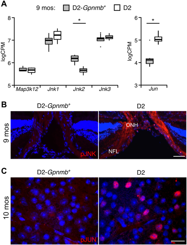
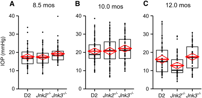
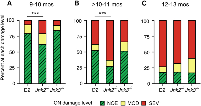
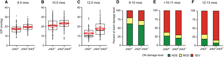
The cJun N-terminal kinases (JNKs; JNK1, JNK2, and JNK3) promote degenerative processes after neuronal injury and in disease. JNK2 and JNK3 have been shown to promote retinal ganglion cell (RGC) death after optic nerve injury. In their absence, long-term survival of RGC somas is significantly increased after mechanical optic nerve injury. In glaucoma, because optic nerve damage is thought to be a major cause of RGC death, JNKs are an important potential target for therapeutic intervention. To assess the role of JNK2 and JNK3 in an ocular hypertensive model of glaucoma, null alleles of Jnk2 and Jnk3 were backcrossed into the DBA/2J (D2) mouse. JNK activation occurred in RGCs following increased intraocular pressure in D2 mice. However, deficiency of both Jnk2 and Jnk3 together did not lessen optic nerve damage or RGC death. These results differentiate the molecular pathways controlling cell death in ocular hypertensive glaucoma compared with mechanical optic nerve injury. It is further shown that JUN, a pro-death component of the JNK pathway in RGCs, can be activated in glaucoma in the absence of JNK2 and JNK3. This implicates JNK1 in glaucomatous RGC death. Unexpectedly, at younger ages, Jnk2-deficient mice were more likely to develop features of glaucomatous neurodegeneration than D2 mice expressing Jnk2. This appears to be due to a neuroprotective effect of JNK2 and not due to a change in intraocular pressure. The Jnk2-deficient context also unmasked a lesser role for Jnk3 in glaucoma. Jnk2 and Jnk3 double knockout mice had a modestly increased risk of neurodegeneration compared with mice only deficient in Jnk2. Overall, these findings are consistent with pleiotropic effects of JNK isoforms in glaucoma and suggest caution is warranted when using JNK inhibitors to treat chronic neurodegenerative conditions.


Conflict of interest statement

The authors declare that they have no conflict of interest.

Figures

Fig. 1
Fig. 1. JNK pathway genes associated with axon injury in RGCs are expressed and activated during D2 glaucoma at 9 months of age.
a Publically available RNA-sequencing data sets from RGCs were assessed for expression of a subset of JNK signaling pathway genes implicated in axon injury. RGCs from a group of D2 eyes with ocular hypertension, but not optic nerve damage were compared with RGCs from control D2-Gpnmb+ eyes that do not have high intraocular pressure or develop glaucoma. The D2 eyes analyzed here belong to the group of eyes with the earliest detected changes in this model (Group 2,). The Tukey-style box plots were generated in R. The thick bar represents median expression level and the hinges correspond to the first and third quartiles. Whiskers represent 1.5 × the interquartile range, with outlying data points plotted individually. Expression level is shown as log counts per million (log2CPM). Significant differences in expression of Jnk2 and Jun were detected. b Phosphorylated S63 pan-JNK immunoreactivity (pJNK, red) in ocular hypertensive (D2) and normotensive (D2-Gpnmb+) eyes was assessed by fluorescence in cross-sections that include the retina and the optic nerve head (ONH). The nerve fiber layer (NFL) is filled by RGC axons as they extend toward the ONH. DAPI (blue) labeling of nuclei was used as a counterstain. c Phosphorylated JUN immunoreactivity (pJUN, red) in the ganglion cell layer was assessed by fluorescence in retinal flat mounts with DAPI (blue) used as a counterstain. Representative images from ocular hypertensive (D2) and normotensive (D2-Gpnmb+) eyes are shown. *FDR < 0.05. Scale bar: 50 μm b, 25 μm c
Fig. 2
Fig. 2. Jnk genotype has no affect on iris disease.
Images of eyes were taken using a slit lamp biomicroscope. a Representative broad beam images demonstrating similar iris abnormalities in mice of all genotypes. b Representative images demonstrating the similar transillumination defects resulting from iris depigmentation in D2, D2.Jnk2−/−, and D2.Jnk3−/− mice
Fig. 3
Fig. 3. Deficiency of Jnk2 or Jnk3 does not alter intraocular pressure profiles in D2 mice.
IOP elevation occurs in Jnk2 and Jnk3-deficient mice at 8.5 mos a, 10.0 mos b, and 12.0 mos c. IOP elevations sufficient to cause glaucoma were observed in mice of all genotypes. No significant differences were found when comparing Jnk-deficient with control D2 mice (n = 40 per group, P> 0.05 in each comparison. However, at 12 mos Jnk2-deficient mice tended to have lower IOP than controls (n = 42, P = 0.052). IOP levels start to decline in D2 mice at 12 mos due to ciliary body degeneration and decreased production of aqueous humor. These data do not exclude a role for JNK2 in lessening this late disease process in the ciliary body, but this IOP difference may also be due to chance variation. In the boxplot, the boxes define the 25th and 75th percentiles. The black line in each box represents the median value. The red diamonds indicate the mean value and the 95% CI. The full range of the data points is also depicted
Fig. 4
Fig. 4. Histological assessment of retina and optic nerve.
Representative images from H&E-stained ocular cross-sections and PPD stained optic nerves are shown for unaffected eyes (6 months of age) a, and severely affected eyes (12 months of age) b. These images demonstrate the hallmark degenerative features of glaucoma including RGC layer (GCL) neuron loss in the retina, nerve fiber layer thinning (compare arrowheads in  a and b), optic disc cupping (arrow), and sick and degenerating axons (darkly stained dots), axon loss and glial scarring (asterisks) in the optic nerve. Scale bars: 50 μm (retina), 100 μm (optic nerve head), 25 μm (optic nerve)
Fig. 5
Fig. 5. Jnk2 deficiency increases optic nerve vulnerability to high IOP.
The severity of optic nerve damage was determined using PPD stained cross-sections (see Materials and methods for details) at 9–10 mos a, > 10–11 mos b, and 12–13 mos c. Nerves were determined to have no glaucomatous nerve damage (no or early, NOE—no glaucoma based on nerve damage but called NOE as some of these eyes have early transcriptional changes that precede degeneration), moderate disease (MOD) or severe disease (SEV). An increased number of eyes from Jnk2 mice had glaucomatous nerve damage compared with control D2 mice at 9.5 and 10.5 mos. ***P < 1 × 10−5
Fig. 6
Fig. 6. Jnk3 deficiency subtly exacerbates glaucoma in mice lacking Jnk2.
ac IOP elevations sufficient to cause glaucoma occurred for both genotypes with no significant differences between genotypes at 8 and 10.5 mos. At 12 mos, the difference between Jnk2−/− and Jnk2−/− Jnk3−/− genotypes was due to the IOP decrease observed in Jnk2 only deficient mice (P = 0.042). The IOP levels in mice with combined Jnk2 and 3 deficiencies closely resembled those of wild-type, heterozygous, and Jnk3-deficient mice (compare with Fig. 3c). Thus, the greater decrease in IOP in the Jnk2−/− mice may be due to chance, as it is hard to reconcile why the additional deficiency of Jnk3 in Jnk2−/− Jnk3−/− mutant mice would prevent it. d-f The severity of optic nerve damage was determined using PPD stained cross-sections (see Materials and methods for details). Data for D2.Jnk2−/− mice are the same data as in Fig. 5 as mice of all genotypes were produced in the same litters and examined at the same time
Fig. 7
Fig. 7. JNK2 and JNK3 are not necessary for RGC somal degeneration in glaucoma.
a The JNK target JUN is known to promote RGC apoptosis in D2 glaucoma. Double deficiency of Jnk2 and Jnk3 did not prevent accumulation of phosphorylated JUN (pJUN) immunoreactivity (red) at 10 mos. pJUN was detected by immunofluorescence in retinal flat mounts. b β-Tubulin is an RGC-specific marker in the retina and was used to label RGCs in retinal flat mounts at 12 mos. Representative images show loss of RGCs in eyes with severe optic nerve degeneration compared with healthy (NOE) eyes. Occasional RGCs have higher β-tubulin immunoreactivity (bright cells). c Quantification of β-tubulin-positive cells suggests that Jnk2 and Jnk3 deficiency did not affect somal degeneration of RGCs in eyes with severe optic nerve loss. RGC counts were similar in NOE eyes for all genotypes (represented together as All)

References

    1. Quigley HA, Broman AT. The number of people with glaucoma worldwide in 2010 and 2020. Br. J. Ophthalmol. 2006;90:262–267. doi: 10.1136/bjo.2005.081224. - DOI - PMC - PubMed
    1. Anderson DR, Hendrickson A. Effect of intraocular pressure on rapid axoplasmic transport in monkey optic nerve. Invest. Ophthalmol. 1974;13:771–783. - PubMed
    1. Burgoyne CF, Downs JC, Bellezza AJ, Suh JK, Hart RT. The optic nerve head as a biomechanical structure: a new paradigm for understanding the role of IOP-related stress and strain in the pathophysiology of glaucomatous optic nerve head damage. Prog. Retin. Eye. Res. 2005;24:39–73. doi: 10.1016/j.preteyeres.2004.06.001. - DOI - PubMed
    1. Howell GR, et al. Axons of retinal ganglion cells are insulted in the optic nerve early in DBA/2J glaucoma. J. Cell. Biol. 2007;179:1523–1537. doi: 10.1083/jcb.200706181. - DOI - PMC - PubMed
    1. Quigley HA, Anderson DR. Distribution of axonal transport blockade by acute intraocular pressure elevation in the primate optic nerve head. Invest. Ophthalmol. Vis. Sci. 1977;16:640–644. - PubMed

Publication types

MeSH terms

Substances