. 2018 Oct;25(10):1796-1807.
doi: 10.1038/s41418-018-0143-2. Epub 2018 Jun 13.

Strong and sustained activation of the anticipatory unfolded protein response induces necrotic cell death

Affiliations

Strong and sustained activation of the anticipatory unfolded protein response induces necrotic cell death

Mara Livezey et al. Cell Death Differ. 2018 Oct.

Abstract

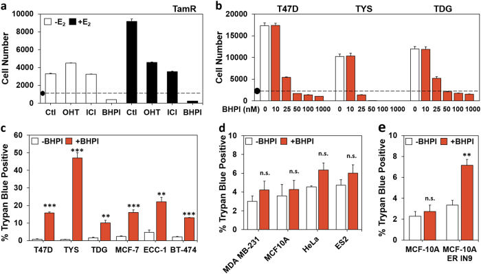
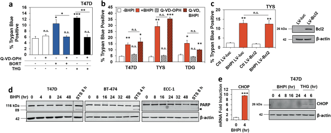
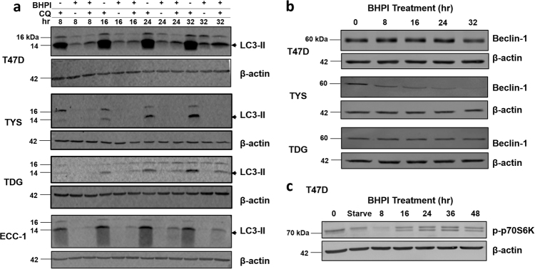
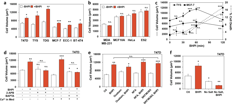
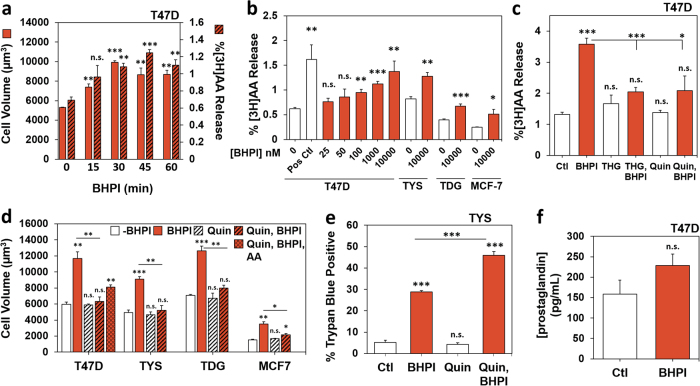
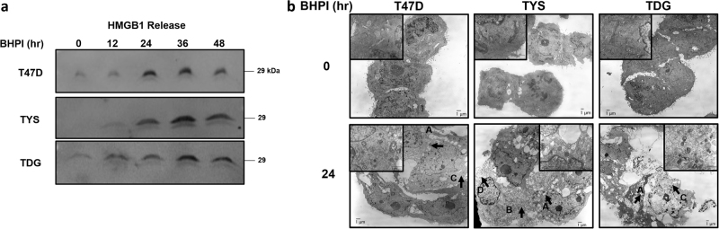
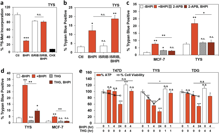
The endoplasmic reticulum stress sensor, the unfolded protein response (UPR), regulates intracellular protein homeostasis. While transient activation of the reactive UPR by unfolded protein is protective, prolonged and sustained activation of the reactive UPR triggers CHOP-mediated apoptosis. In the recently characterized, evolutionarily conserved anticipatory UPR, mitogenic hormones and other effectors pre-activate the UPR; how strong and sustained activation of the anticipatory UPR induces cell death was unknown. To characterize this cell death pathway, we used BHPI, a small molecule that activates the anticipatory UPR through estrogen receptor α (ERα) and induces death of ERα+ cancer cells. We show that sustained activation of the anticipatory UPR by BHPI kills cells by inducing depletion of intracellular ATP, resulting in classical necrosis phenotypes, including plasma membrane disruption and leakage of intracellular contents. Unlike reactive UPR activation, BHPI-induced hyperactivation of the anticipatory UPR does not induce apoptosis or sustained autophagy. BHPI does not induce CHOP protein or PARP cleavage, and two pan-caspase inhibitors, or Bcl2 overexpression, have no effect on BHPI-induced cell death. Moreover, BHPI does not increase expression of autophagy markers, or work through recently identified programmed-necrosis pathways, such as necroptosis. Opening of endoplasmic reticulum IP3R calcium channels stimulates cell swelling, cPLA2 activation, and arachidonic acid release. Notably, cPLA2 activation requires ATP depletion. Importantly, blocking rapid cell swelling or production of arachidonic acid does not prevent necrotic cell death. Rapid cell death is upstream of PERK activation and protein synthesis inhibition, and results from strong and sustained activation of early steps in the anticipatory UPR. Supporting a central role for ATP depletion, reversing ATP depletion blocks rapid cell death, and the onset of necrotic cell death is correlated with ATP depletion. Necrotic cell death initiated by strong and sustained activation of the anticipatory UPR is a newly discovered role of the UPR.


Conflict of interest statement

The authors declare that they have no conflict of interest.

Figures

Fig. 1
Fig. 1
BHPI kills ERα+ breast and endometrial cancer cells. a TamR cell proliferation after 4 days in 8% charcoal:dextran-treated calf (CD-calf) serum with the indicated treatments: E2 100 pM; z-4-hydroxytamoxifen (OHT, the active form of tamoxifen) 1 µM; ICI/fulvestrant 1 µM; BHPI 100 nM. (•) Indicates starting cell number (day 0). b Dose−response study of the effect of increasing concentrations of BHPI on proliferation of T47D, TYS, and TDG cells after 4 days. a, b Alamar Blue assays (n = 8 biological replicate experiments). c Automated trypan blue exclusion assays of ERα-positive cells after 24 h treatment with 1 µM BHPI. d Trypan blue exclusion assays of ERα-negative cells after 24 h treatment with 1 µM BHPI. e Trypan blue exclusion assays of isogenic MCF10A and MCF10AER IN9 cells after 24-h treatment with 1 µM BHPI. ce Data is mean ± s.e.m. (n = 3 biological replicate experiments). For (ce) **p < 0.01, ***p < 0.001, n.s. = not significant by Student’s t test
Fig. 2
Fig. 2
BHPI does not induce caspase-dependent apoptosis. a Trypan blue exclusion assay of T47D cells after 24 h treatment with or without 20 µM Q-VD-OPH, 100 nM bortezomib (BORT), or 10 µM thapsigargin (THG). b Trypan blue exclusion assay of T47D and TDG cells after 24 h, and TYS cells after 1 h treatment with: 1 µM BHPI with or without 20 µM Q-VD-OPH. c Trypan blue exclusion assay and western blot analysis of TYS cells infected with lentivirus expressing luciferase (LV-luc) or Bcl2 (LV-Bcl2) after 1 h treatment with 1 µM BHPI. ac Data are mean ± s.e.m. (n = 3 biological replicate experiments), *p < 0.05, **p < 0.01, ***p < 0.001, n.s. = not significant by Student’s t test. d Western blot analysis of cleaved PARP (arrow) in T47D and BT-474 breast cancer cells, and ECC-1 endometrial cancer cells after treatment with 1 µM BHPI or 5 µM staurosporine (STS) for the indicated times. e mRNA and western blot analysis of CHOP induction in T47D cells treated with 1 µM BHPI or 300 nM thapsigargin (THG) for the indicated times
Fig. 3
Fig. 3
Autophagy is not upregulated by BHPI. a Western blot analysis of LC3-II production (arrow) in T47D, TYS, TDG, and ECC-1 cells after treatment with 1 µM BHPI, 30 µM Chloroquine (CQ), or BHPI + CQ for the indicated times. b Western blot analysis of Beclin-1 levels in T47D, TYS, and TDG cells after treatment with 1 µM BHPI for the indicated times. c Western blot analysis of phosphorylation of p70S6K in T47D cells after treatment with 1 µM BHPI for the indicated times, or after serum starvation (Starve) for 24 h
Fig. 4
Fig. 4
BHPI-ERα-induced cell swelling is linked to early steps in the anticipatory UPR. a Cell volume in ERα-positive cells treated with 1 µM BHPI for 1 h (T47D, TYS, TDG, MCF-7, and BT-474), or 30 min (ECC-1). b Cell volume in ERα-negative MDA MB-231, MCF10A, HeLa, and ES2 cells treated with 1 µM BHPI for 1 h. c Time course of the effect of BHPI on cell volume and cell death in TYS and MCF-7 cells. Cell volume (solid lines) and cell death (dashed lines) were assayed by trypan blue exclusion in cells treated with 50 nM BHPI (TYS), or 100 nM BHPI (MCF-7). d Blocking the increase in intracellular calcium with 2-APB, or with the calcium chelator, BAPTA-AM (BAPTA), prevents the BHPI-induced increase in cell volume. Cell volume in T47D cells treated for 1 h with vehicle, 50 nM BHPI, 100 µM 2-APB, or BHPI + 2-APB in calcium-free isosmotic medium, or vehicle, 20 µM BAPTA-AM, or BHPI + BAPTA-AM. e, f Cell volume of T47D cells after 1 h treatment as indicated. e BHPI 50 nM; ouabain 2 µM (Na+/K+ ATPase inhibitor); niflumic acid (NFA) 100 µM (nonspecific anion channel inhibitor); SKF96365 10 µM (TRP channel inhibitor). f BHPI 50 nM; isosmotic salt-free sucrose buffer (No Salt). Data are mean ± s.e.m. (n = 3 biological replicate experiments) *p < 0.05, **p < 0.01, ***p < 0.001, n.s. = not significant by Student’s t test
Fig. 5
Fig. 5
BHPI activation of cPLA2 causes release of arachidonic acid (AA). a Time course showing a correlation between increased AA release and increased cell volume in BHPI-treated T47D cells. Time course of changes in cell volume (solid bars) and 3H-arachidonic acid release ([3H]AA, hatched bars) after treatment of T47D cells with vehicle or with 10 µM BHPI. Data are mean ± s.e.m. (solid bars, n = 3 biological replicate experiments; hatched bars, n = 4 biological replicate experiments). b Concentration-dependent release of [3H]AA from T47D, TYS, TDG, and MCF-7 cell membranes after treatment for 45 min with vehicle or the indicated concentration of BHPI. Positive control (Pos Ctl) is 150 mOsm buffer with 1 µM THG. c THG and the cPLA2 inhibitor quinacrine (Quin) block the BHPI-mediated increase in AA. Release of [3H]AA from T47D cell membranes after treatment for 45 min with vehicle, 1 µM thapsigargin (THG), 1 µM BHPI, BHPI + THG, 300 µM Quin, or BHPI + Quin. d Effect of Quin and AA on BHPI-induced changes in cell volume. Volume of T47D, TYS, TDG, and MCF-7 cells after treatment for 1 h with vehicle, 50 nM BHPI, 300 µM Quin, 250 µM AA, BHPI + Quin, or BHPI + Quin + AA. e Quin does not block BHPI-induced cell death. Trypan blue exclusion assay for cell death in TYS cells treated for 1 h with vehicle, 1 µM BHPI, 300 µM Quin, or BHPI + Quin. f ELISA determination of prostaglandin production from T47D cells after treatment for 24 h with 10 µM BHPI. Data are mean ± s.e.m. b, c, f (n = 4 biological replicate experiments), d, e (n = 3 biological replicate experiments). For (af) *p < 0.05, **p < 0.01, ***p < 0.001, n.s. = not significant by Student’s t test
Fig. 6
Fig. 6
BHPI-induced cell death exhibits a necrotic phenotype. a Western blot analysis of HMGB1 release into the cell culture supernatant at the indicated time points from T47D, TYS, and TDG cells treated with 1 µM BHPI. Equal volumes of culture supernatant were loaded for analysis. b Transmission electron microscopy (TEM) images of T47D, TYS, and TDG cells treated with 1 µM BHPI for 0 or 24 h. Letters with arrows indicate features characteristic of necrotic morphology: a swollen vacuoles, b swollen mitochondria, c cytoplasmic lightening, d membrane rupture/“ghost” cells
Fig. 7
Fig. 7
BHPI-induced necrosis is an upstream effect of anticipatory UPR activation and requires ATP depletion. a Incorporation of 35S-methionine into newly synthesized protein in TYS cells treated for 1 h with vehicle (set to 100%), 50 nM BHPI, 200 nM ISRIB, BHPI + ISRIB, or 10 µM cyclohexamide (CHX). b ISRIB does not prevent BHPI-induced cell death. Trypan blue exclusion assay for cell death in TYS cells after 1 h treatment with vehicle, 1 µM BHPI, 200 nM ISRIB, or BHPI + ISRIB. c Locking the EnR IP3R calcium channels closed with 2-APB blocks BHPI-induced cell death. Trypan blue exclusion assay for cell death in MCF-7 and TYS cells treated for 1 h with vehicle, 1 µM BHPI, 100 µM 2-APB, or BHPI + 2-APB. d THG inhibits BHPI-induced cell death. Trypan blue exclusion assay for cell death in TYS and MCF-7 cells treated for 1 h with vehicle, 1 µM BHPI, 10 µM THG, or BHPI + THG. e Time-dependent decline in ATP levels in BHPI-treated cells correlates with cell death. Measurement of whole-cell ATP levels (bars) and trypan blue exclusion (overlay, [ο]) after treatment for the indicated times with vehicle, 1 µM BHPI, 10 µM THG, or BHPI + THG. For TYS cells, ~23% of cells have died at early times and disintegrated at 24 h and are not counted by the instrument. Therefore, the percentage of trypan blue excluding cells declines at 24 h, the total number of dead cells (trypan blue positive + disintegrated cells) is estimated to be ~59%. Data are mean ± s.e.m. a (n = 4 biological replicate experiments), bd (n = 3 biological replicate experiments), e (n = 5 biological replicate experiments; ATP) (n = 3 biological replicate experiments; cell viability) *p < 0.05, **p < 0.01, ***p < 0.001, n.s. = not significant by Student’s t test

References

    1. Walter P, Ron D. The unfolded protein response: from stress pathway to homeostatic regulation. Science. 2011;334:1081–6. doi: 10.1126/science.1209038. - DOI - PubMed
    1. Ma Y, Hendershot LM. The role of the unfolded protein response in tumour development: friend or foe? Nat Rev Cancer. 2004;4:966–77. doi: 10.1038/nrc1505. - DOI - PubMed
    1. Wang M, Kaufman RJ. The impact of the endoplasmic reticulum protein-folding environment on cancer development. Nat Rev Cancer. 2014;14:581–97. doi: 10.1038/nrc3800. - DOI - PubMed
    1. Lee AS. GRP78 induction in cancer: therapeutic and prognostic implications. Cancer Res. 2007;67:3496–9. doi: 10.1158/0008-5472.CAN-07-0325. - DOI - PubMed
    1. Urra H, Dufey E, Avril T, Chevet E, Hetz C. Endoplasmic reticulum stress and the hallmarks of cancer. Trends Cancer. 2016;2:252–62. doi: 10.1016/j.trecan.2016.03.007. - DOI - PubMed

Publication types

MeSH terms