

Clinical features, fungal load, coinfections, histological skin changes, and itraconazole treatment response of cats with sporotrichosis caused by Sporothrix brasiliensis
- PMID: 29899416
- PMCID: PMC5998065
- DOI: 10.1038/s41598-018-27447-5
Clinical features, fungal load, coinfections, histological skin changes, and itraconazole treatment response of cats with sporotrichosis caused by Sporothrix brasiliensis
Abstract
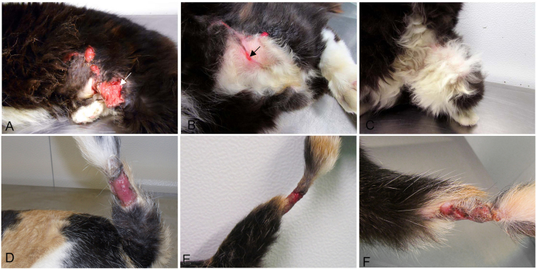
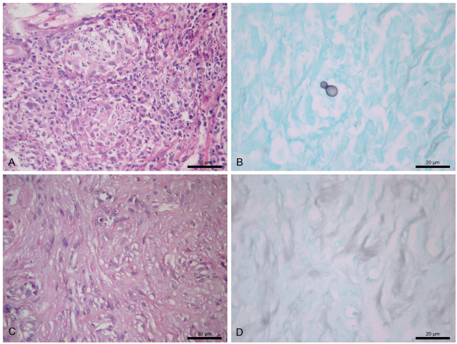
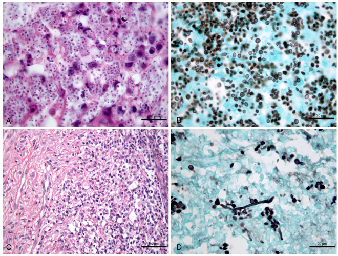
Zoonotic sporotrichosis caused by the fungus Sporothrix brasiliensis is usually severe in cats. This study investigated the associations between clinical features, fungal load, coinfections, histological skin changes, and response to itraconazole in cats with sporotrichosis caused by S. brasiliensis. Fifty-two cats with skin lesions and a definitive diagnosis of sporotrichosis were treated with itraconazole for a maximum period of 36 weeks. The animals were submitted to clinical examination and two subsequent collections of samples from the same skin lesion for fungal diagnosis and histopathology, as well as serology for feline immunodeficiency (FIV) and leukaemia (FeLV) viruses. Thirty-seven (71%) cats were clinically cured. Nasal mucosa lesions and respiratory signs were associated with treatment failure. Cats coinfected with FIV/FeLV (n = 12) had a lower neutrophil count in the lesion. A high fungal load in skin lesions was linked to young age and treatment failure, as well as to a longer time of wound healing, poorly formed granulomas and fewer neutrophils, macrophages and lymphocytes in these lesions. These results indicate that itraconazole is effective, but nasal mucosal involvement, respiratory signs and high fungal loads in skin lesions are predictors of treatment failure that will assist in the development of better treatment protocols for cats.
Conflict of interest statement
The authors declare no competing interests.
Figures
Similar articles
-
Feline sporotrichosis in Asia.Braz J Microbiol. 2021 Mar;52(1):125-134. doi: 10.1007/s42770-020-00274-5. Epub 2020 May 3. Braz J Microbiol. 2021. PMID: 32363567 Free PMC article. Review.
-
Co-infection with feline retrovirus is related to changes in immunological parameters of cats with sporotrichosis.PLoS One. 2018 Nov 30;13(11):e0207644. doi: 10.1371/journal.pone.0207644. eCollection 2018. PLoS One. 2018. PMID: 30500849 Free PMC article.
-
Feline sporotrichosis: a case series of itraconazole-resistant Sporothrix brasiliensis infection.Braz J Microbiol. 2021 Mar;52(1):163-171. doi: 10.1007/s42770-020-00290-5. Epub 2020 May 9. Braz J Microbiol. 2021. PMID: 32388779 Free PMC article.
-
Sporothrix brasiliensis Treatment Failure without Initial Elevated Itraconazole MICs in Felids at Border of Brazil.Emerg Infect Dis. 2025 Sep;31(9):1783-1792. doi: 10.3201/eid3109.250156. Emerg Infect Dis. 2025. PMID: 40867021
-
Guideline for the management of feline sporotrichosis caused by Sporothrix brasiliensis and literature revision.Braz J Microbiol. 2021 Mar;52(1):107-124. doi: 10.1007/s42770-020-00365-3. Epub 2020 Sep 29. Braz J Microbiol. 2021. PMID: 32990922 Free PMC article. Review.
Cited by
-
Sporothrix Brasiliensis: A Review of an Emerging South American Fungal Pathogen, Its Related Disease, Presentation and Spread in Argentina.J Fungi (Basel). 2021 Feb 26;7(3):170. doi: 10.3390/jof7030170. J Fungi (Basel). 2021. PMID: 33652625 Free PMC article. Review.
-
Feline sporotrichosis in Asia.Braz J Microbiol. 2021 Mar;52(1):125-134. doi: 10.1007/s42770-020-00274-5. Epub 2020 May 3. Braz J Microbiol. 2021. PMID: 32363567 Free PMC article. Review.
-
Application of Principal Component Analysis as a Prediction Model for Feline Sporotrichosis.Vet Sci. 2025 Jan 9;12(1):32. doi: 10.3390/vetsci12010032. Vet Sci. 2025. PMID: 39852907 Free PMC article.
-
Phenotypic and Genotypic Characterization and Antifungal Susceptibility of Sporothrix schenckii sensu stricto Isolated from a Feline Sporotrichosis Outbreak in Bangkok, Thailand.J Fungi (Basel). 2023 May 18;9(5):590. doi: 10.3390/jof9050590. J Fungi (Basel). 2023. PMID: 37233301 Free PMC article.
-
Trends in Molecular Diagnostics and Genotyping Tools Applied for Emerging Sporothrix Species.J Fungi (Basel). 2022 Jul 31;8(8):809. doi: 10.3390/jof8080809. J Fungi (Basel). 2022. PMID: 36012797 Free PMC article. Review.
References
Publication types
MeSH terms
Substances
LinkOut - more resources
Full Text Sources
Other Literature Sources
Miscellaneous
