. 2018 Jun;558(7710):435-439.
doi: 10.1038/s41586-018-0218-8. Epub 2018 Jun 13.

Quantitative phosphoproteomic analysis of the molecular substrates of sleep need

Affiliations

Quantitative phosphoproteomic analysis of the molecular substrates of sleep need

Zhiqiang Wang et al. Nature. 2018 Jun.

Abstract

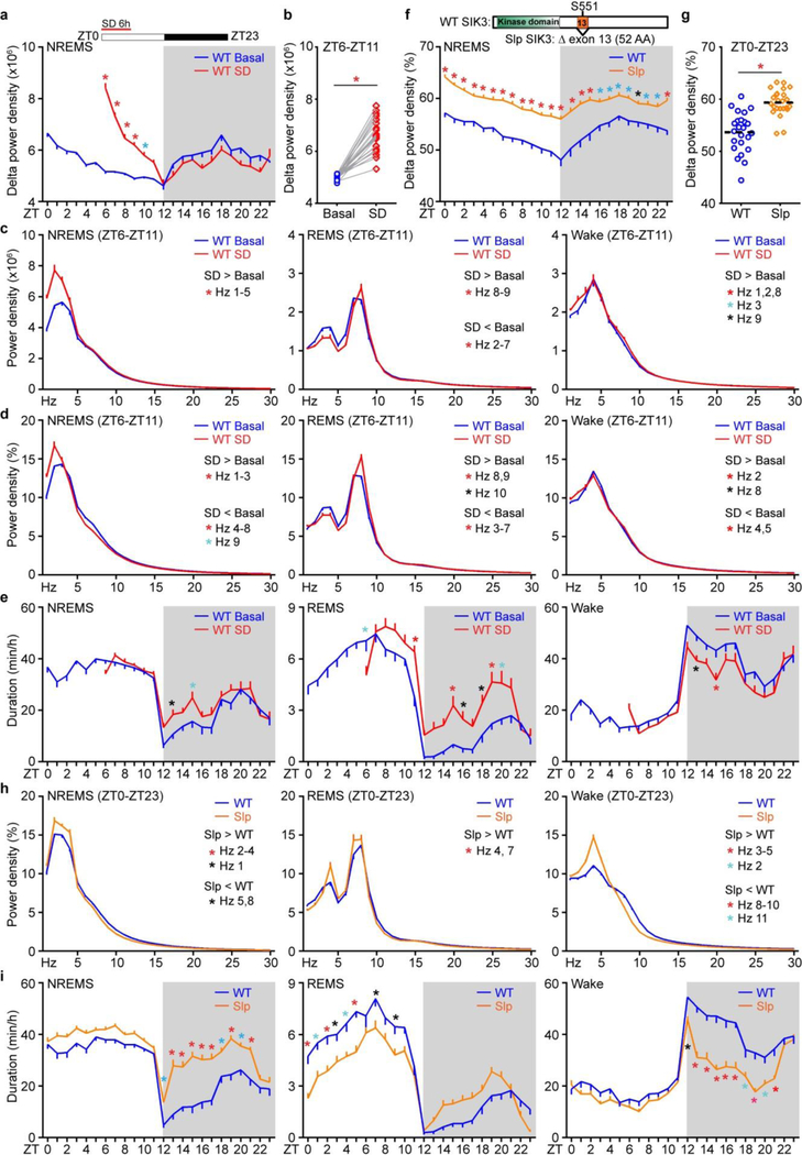
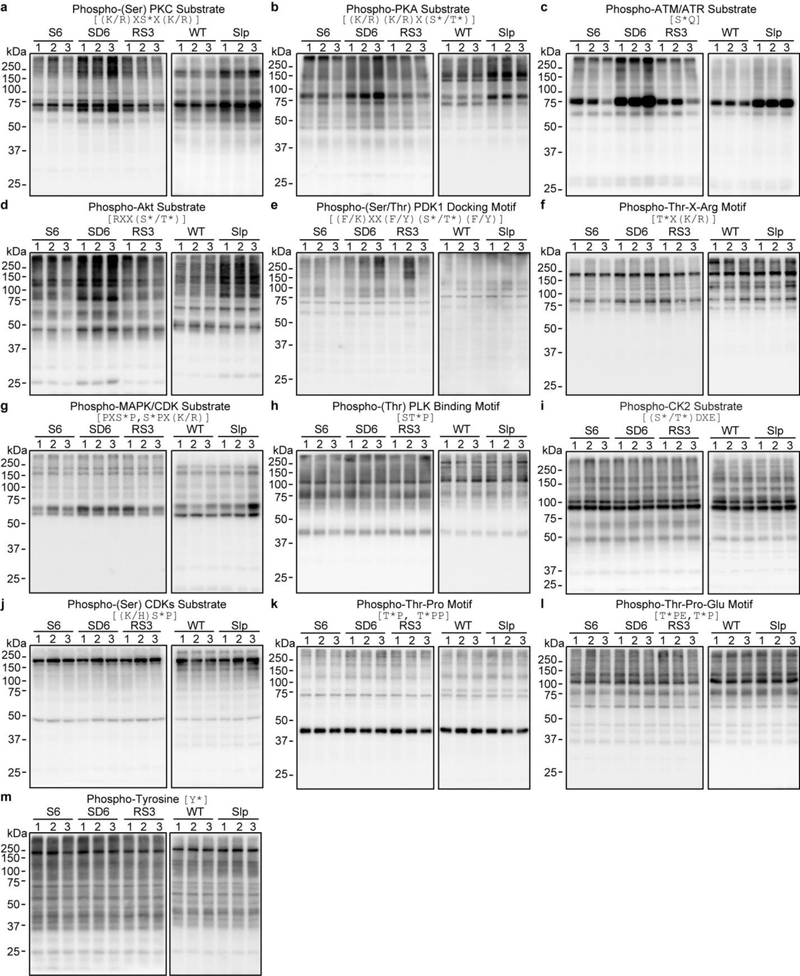
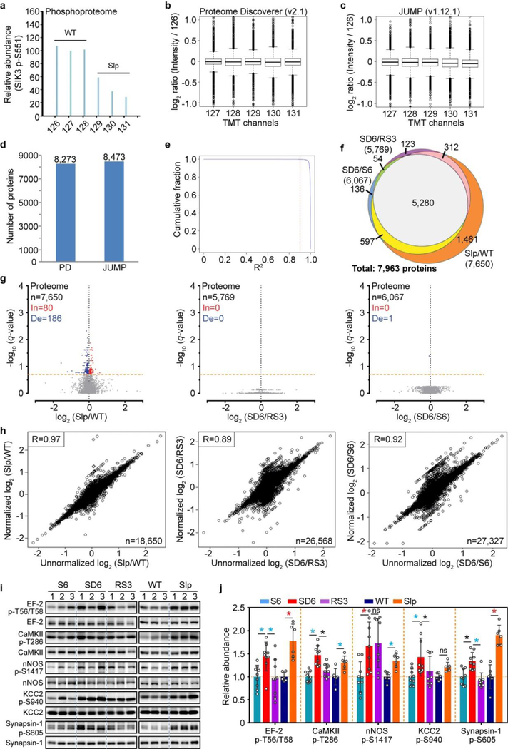
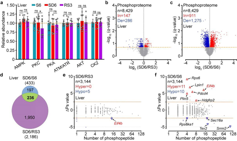
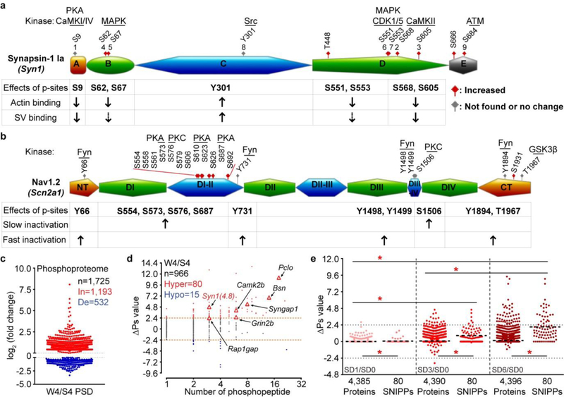
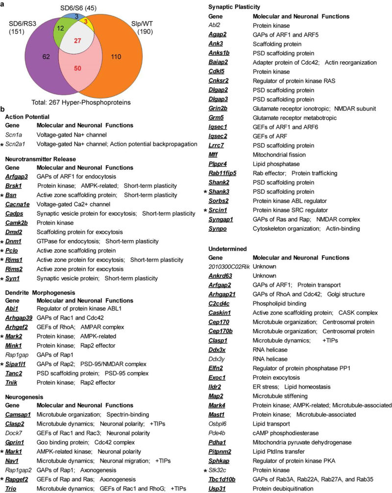
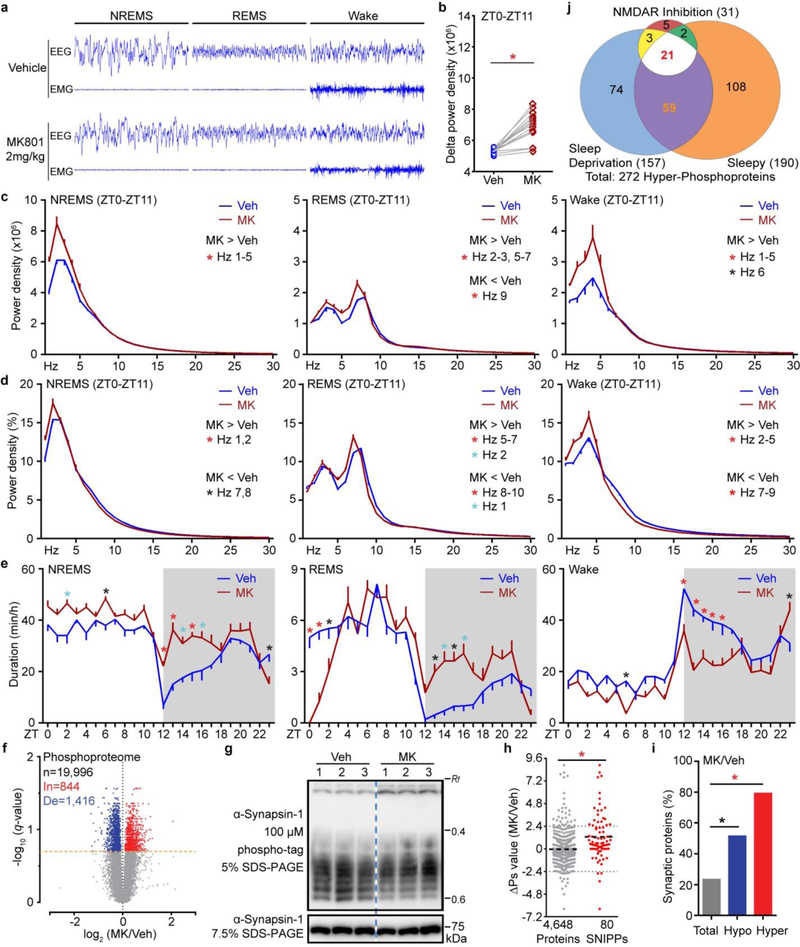
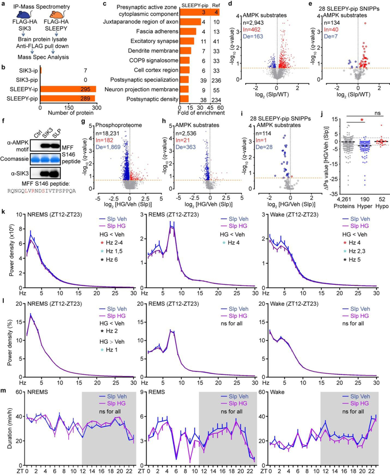
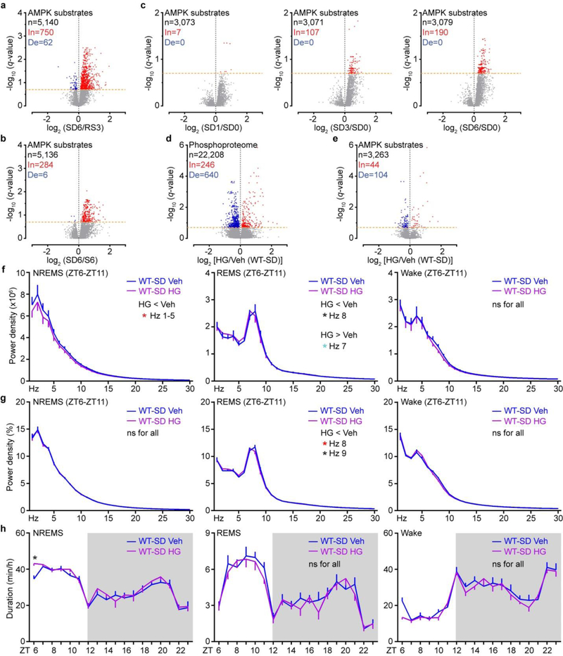
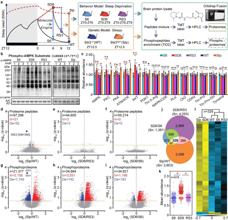
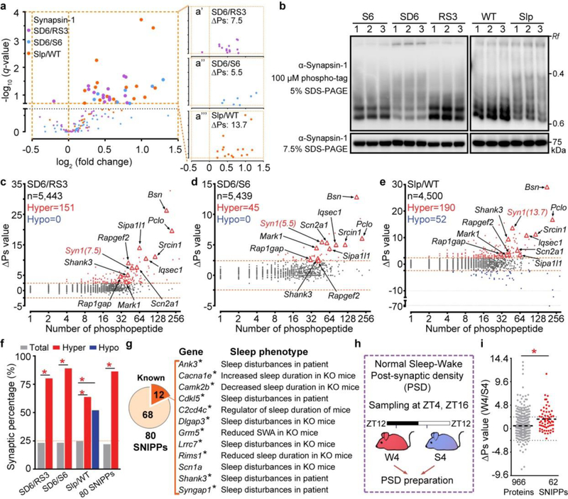
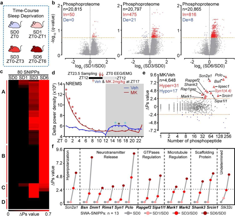
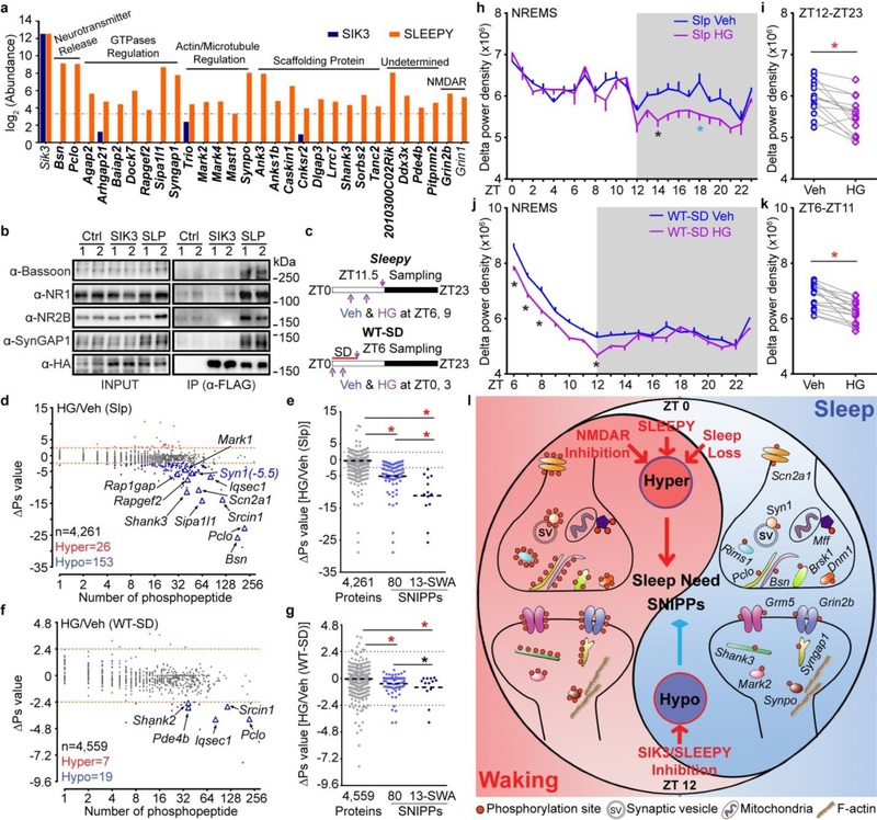
Sleep and wake have global effects on brain physiology, from molecular changes1-4 and neuronal activities to synaptic plasticity3-7. Sleep-wake homeostasis is maintained by the generation of a sleep need that accumulates during waking and dissipates during sleep8-11. Here we investigate the molecular basis of sleep need using quantitative phosphoproteomic analysis of the sleep-deprived and Sleepy mouse models of increased sleep need. Sleep deprivation induces cumulative phosphorylation of the brain proteome, which dissipates during sleep. Sleepy mice, owing to a gain-of-function mutation in the Sik3 gene 12 , have a constitutively high sleep need despite increased sleep amount. The brain proteome of these mice exhibits hyperphosphorylation, similar to that seen in the brain of sleep-deprived mice. Comparison of the two models identifies 80 mostly synaptic sleep-need-index phosphoproteins (SNIPPs), in which phosphorylation states closely parallel changes of sleep need. SLEEPY, the mutant SIK3 protein, preferentially associates with and phosphorylates SNIPPs. Inhibition of SIK3 activity reduces phosphorylation of SNIPPs and slow wave activity during non-rapid-eye-movement sleep, the best known measurable index of sleep need, in both Sleepy mice and sleep-deprived wild-type mice. Our results suggest that phosphorylation of SNIPPs accumulates and dissipates in relation to sleep need, and therefore SNIPP phosphorylation is a molecular signature of sleep need. Whereas waking encodes memories by potentiating synapses, sleep consolidates memories and restores synaptic homeostasis by globally downscaling excitatory synapses4-6. Thus, the phosphorylation-dephosphorylation cycle of SNIPPs may represent a major regulatory mechanism that underlies both synaptic homeostasis and sleep-wake homeostasis.


Conflict of interest statement

The authors declare no competing financial interests.

Figures

Extended Data Figure 1 |
Extended Data Figure 1 |. Sleep phenotype analysis of the sleep-deprived and Sleepy models.
a-e, Analysis of circadian (a) and mean (b) absolute NREMS delta power, absolute EEG power spectra (c), relative EEG power spectra (d) and duration (e) of NREMS, REMS, wake states of wild-type mice (n = 24) without (WT Basal) and with 6-h sleep deprivation (WT SD). f-i, Analysis of circadian (f) and mean (g) relative NREMS delta power, relative EEG power spectra (h) and duration (i) of NREMS, REMS, wake states of Sik3+/+ (WT, n = 24) and Sik3Slp/+ (Slp, n = 24) mice. Mean ± s.e.m., two-way ANOVA with Sidak’s test (a, c-f, h, i); Paired t-test, two-tailed (b); Mean, unpaired t-test, two-tailed (g). *(black) P < 0.05; *(cyan) P < 0.01; *(red) P < 0.001.
Extended Data Figure 2 |
Extended Data Figure 2 |. Analysis of global signaling changes in two models of increased sleep need.
a-m, Representative immunoblots of 13 phospho-motif antibodies to assess global signaling changes in whole brain lysates of two models [three (sleep-deprived) or two (Sleepy) independent experiments]. Quantitative analysis of immunoblots of all 14 phospho-motif antibodies is shown in Figure 1c [n=12 (S6), 9 (SD6, RS3), 6 (WT, Slp)].
Extended Data Figure 3 |
Extended Data Figure 3 |. Analysis of sleep phenotype and signaling changes after food/water deprivation in the baseline and sleep deprivation conditions.
a-c, Analysis of circadian (a) and mean (b) absolute NREMS delta power, duration (c) of NREMS, REMS, wake states of wild-type mice (n = 8) without (Sham) or with 6-h food/water deprivation (FD 6h). d, Quantitative analysis of immunoblots with 7 phospho-motif antibodies using whole brain lysates of sham and FD 6h mice (n = 8) harvested at ZT6. e-g, Analysis of circadian (e) and mean (f) absolute NREMS delta power, duration (g) of NREMS, REMS, wake states of wild-type mice (n = 11) without (SD+Sham) or with 6-h food/water deprivation during 6-h sleep deprivation (SD+FD 6h). h, Quantitative analysis of immunoblots with 7 phospho-motif antibodies using whole brain lysates of SD+Sham and SD+FD mice (n = 6) harvested at ZT6. Mean ± s.e.m., two-way ANOVA, Sidak’s test (a, c, e, g); Paired t-test, two-tailed (b, f); Mean ± s.d., two-way ANOVA, Fisher’s LSD test (d, h). *(black) P < 0.05; ns, P > 0.05.
Extended Data Figure 4 |
Extended Data Figure 4 |. Quality assessment of proteomic and phosphoproteomic analysis.
a, Representative TMT quantification spectrum for the p-S551 containing phosphopeptide from the skipped Sik3 exon-13 among phosphoproteomic data of the Sleepy model (two independent experiments). b-e, Quality assessment of one proteomic dataset (EX4, SlpWTpa2) by two search pipelines. Global distribution of protein quantification using Proteome Discoverer (PD v2.1; n = 8,273) (b) and JUMP (v1.12.1; n = 8,473). Boxes correspond to the 25th, 50th, and 75th percentiles of the data, whiskers extend 1.5-fold of the interquartile range. (c). A similar number of accepted proteins (1% FDR) were identified by two pipelines (d). Pearson correlation between the two pipelines was calculated for each PSM from quantified proteins by both pipelines (e). The vast majority (99.88%) of PSMs (n = 73,454) have R2 larger than 0.9 (red dashed line). f, A Venn diagram showing overlaps of quantified proteins between whole brain proteomes of Sleepy and sleep-deprived models. g, Volcano plots showing comparative analysis of Slp/WT, SD6/RS3 and SD6/S6 proteomes. Multiple unpaired t-test (p-value) following FDR (q-value) analysis. X-axis, log2 (fold change) in abundance; Y-axis, -log10 (q-value) of abundance change. The numbers of total (n), increased [In: q < 0.2, red] and decreased [De: q < 0.2, blue] subjects are shown. Orange dotted lines (q = 0.2). h, Pearson correlation between normalized and unnormalized phosphopeptides in Slp/WT, SD6/RS3, SD6/S6 groups. The numbers of phosphopeptides that can be normalized are shown. i, Immunoblots were performed with phospho-site specific antibodies to verify hyper-phosphorylation of several proteins in two models [three (sleep-deprived) or two (Sleepy) independent experiments]. j, Quantitative analysis of immunoblots using phospho-site specific antibodies (i), normalized with whole protein abundance, for Sleepy (n = 6) and sleep-deprived (n = 9) models. Mean ± s.d., two-way ANOVA with Fisher’s LSD test. *(black) P < 0.05; *(cyan) P < 0.01; *(red) P < 0.001; ns, P > 0.05.
Extended Data Figure 5 |
Extended Data Figure 5 |. Liver phosphoproteome analysis of the sleep-deprived model.
a, Quantitative analysis of immunoblots with 7 phospho-motif antibodies using whole liver lysates from the sleep-deprived model. n =8 (S6), 10 (SD6), 7 (RS3); Mean ± s.d., two-way ANOVA with Fisher’s LSD test. *(black) P < 0.05; ns, P > 0.05. b-c, Volcano plots showing comparative analysis of liver phosphoproteomes in the SD6/RS3 (b) and SD6/S6 (c) groups. Multiple unpaired t-test (p-value) following FDR (q-value) analysis. X-axis, log2 (fold change) in abundance; Y-axis, -log10 (q-value) of abundance change. The numbers of total (n), increased [In: q < 0.2, red] and decreased [De: q < 0.2, blue] subjects are shown. Orange dotted lines (q = 0.2). d, A Venn diagram showing overlaps of significant changed (q < 0.2) phosphopeptides among the SD6/RS3 and SD6/S6 groups. e-f, Global ∆Ps analysis of all phosphoproteins identified in the SD6/RS3 (e) and SD6/S6 (f) groups of liver phosphoproteomes. Dotted lines (∆Ps = +/−2.4).
Extended Data Figure 6 |
Extended Data Figure 6 |. Examples of cumulative phosphorylation of SNIPPs and synaptic phosphoproteomic analysis of normal sleep-wake model.
a-b, A schematic diagram of the domain structure of Synapsin-1 (a) and Nav1.2,, (b) that summarizes known phosphorylation sites, kinases and physiological functions. Synapsin-1 can be divided into five domains (domain A-E). Nav1.2 can be divided into cytoplasmic N-terminal (NT), C-terminal (CT), four homologous transmembrane domains (DI-DIV) and intracellular loops (DI-II, DII-III, DIII-IV). Amino acid numbers refer to the sequence of mouse proteins. Sites 1–9 of Synapsin-1 are designated according to the consensus in the literature. While undetected or unchanged phosophorylation sites are labeled in grey, significantly increased phosphorylation sites are in red. Dashed arrows indicate the presence of contrasting data for biological functions in the literature. c, Published forebrain PSD phosphoproteome results [Diering et al. TableS2] were used for comparative analysis between normal slept (S4) and wake (W4) brains. d, Global ∆Ps analysis of all identified phosphoproteins in the W4/S4 group. Dotted lines (∆Ps = +/−2.4). e, Quantitative ∆Ps analysis of SD1/SD0, SD3/SD0, SD6/SD0 groups. Mean; one-way ANOVA, Tukey’s (Total, SNIPPs); Unpaired t-test, two-tailed (Total vs. SNIPPs). *(red) P < 0.001.
Extended Data Figure 7 |
Extended Data Figure 7 |. Physiological functions of 80 SNIPPs.
a, A Venn diagram showing overlaps of the Hyper-phosphoproteins (∆Ps > 2.4) between sleep-deprived and Sleepy models. b, A summary of 80 SNIPPs and their physiological functions. Stars mark the 13 SWA-SNIPPs (Fig. 3f). Gene names for annotated synaptic proteins are shown in bold.
Extended Data Figure 8 |
Extended Data Figure 8 |. Phospho-state changes of SNIPPs correspond to changes of sleep need in NMDAR inhibition model.
a, Representative 8-s EEG and EMG from ZT0-ZT3 for NREMS, REMS and wake of vehicle and MK801-treated mice. b, Mean absolute NREMS delta power analysis of Veh- or MK-injected mice (n = 14). Paired t-test, two-tailed. c-e, Analysis of absolute EEG power spectra (c), relative EEG power spectra (d) and duration (e) of Veh- or MK-injected wild-type mice (n = 14). Mean ± s.e.m., two-way ANOVA with Sidak’s test. f, Volcano plot showing MK/Veh phosphoproteome comparison. Orange dotted line (q = 0.2). Multiple unpaired t-test (p-value) following FDR (q-value) analysis. g, Phosphorylation state of Synapsin-1 was assessed by phospho-tag (top) and regular (bottom) SDS-PAGE followed by immunoblotting with anti-Synapsin-1 antibody. The Rf value of 1.0 is defined as the position of bromphenol blue dye (two independent experiments). h, Quantitative ∆Ps analysis of MK/Veh group. Mean, unpaired t-test, two-tailed. i, Percentage of synaptic proteins among the total, Hypo- and Hyper-phosphoproteins in MK/Veh group. Chi-square test, two-sided. j, Venn diagram showing overlaps of Hyper-phosphoproteins (∆Ps > 2.4) among all three (Sleepy, SD and MK) models. *(black) P < 0.05; *(cyan) P < 0.01; *(red) P < 0.001.
Extended Data Figure 9 |
Extended Data Figure 9 |. SLEEPY causes constitutively high sleep need by preferentially associating with and phosphorylating SNIPPs.
a, Experimental design for comparing the interactomes of SIK3 and SLEEPY from whole brain lysates. b, Summary of SIK3 and SLEEPY interacting proteins (ip) and preferential interacting proteins (pip). c, Gene-annotation enrichment analysis of 289 SLEEPY preferential interacting proteins (SLEEPY-pip). GO cellular component enrichment analysis using all 22,262 genes of Mus musculus as reference (Ref). Fisher’s Exact with FDR multiple test correction was used to determine statistical significance. Top 10 GO terms of fold enrichment (FDR < 0.0001), the gene number of SLEEPY-pip and Ref in each term are shown. d-e, Volcano plots showing phosphorylation changes of all putative AMPK substrates in the Slp/WT group (d) or from the 28 SLEEPY-pip SNIPPs (e). Orange dotted lines (q = 0.2). f, In vitro kinase assay of recombinant SLEEPY and SIK3, and immunoblotting with AMPK phospho-motif antibody (two independent experiments). g–i, Volcano plot showing comparative analysis of whole brain phosphoproteomes (g), all putative AMPK substrates (h) or from 28 SLEEPY-pip SNIPPs (i) in the HG/Veh (Slp) group. Orange dotted lines (q = 0.2). j, Quantitative ∆Ps analysis of 190 Hyper-phosphoproteins and 52 Hypo-phosphoproteins in HG/Veh (Slp) group. Dotted lines (∆Ps = +/−2.4). k-m, Analysis of absolute EEG power spectra (k), relative EEG power spectra (l), duration (m) of NREMS, REMS and wake states of Sik3Slp/+ (Slp, n = 14) mice injected with vehicle (Veh) or 8mg/kg HG-9–91-01 (HG) at ZT6 and ZT9. Multiple unpaired t-test (p-value) following FDR (q-value) analysis (d-e, g-i). Mean, one-way ANOVA with Dunnett’s test (j). Mean ± s.e.m., two-way ANOVA with Sidak’s test (k-m). *(black) P < 0.05; *(cyan) P < 0.01; *(red) P < 0.001; ns, P > 0.05.
Extended Data Figure 10 |
Extended Data Figure 10 |. Inhibition of SIK3 kinase activity reduced phosphorylation of AMPK substrates in sleep-deprived wild-type brains.
a-c, Volcano plots showing phosphorylation changes of all putative AMPK substrates in the SD6/RS3 (a), SD6/S6 (b) and time-course sleep-deprivation groups (c). Orange dotted lines (q = 0.2). d-e, Volcano plots showing comparative analysis of whole brain phosphoproteome (d) and phosphorylation changes of all putative AMPK substrates (e) in the HG/Veh (WT-SD) group. Orange dotted lines (q = 0.2). f-h, Analysis of absolute EEG power spectra (f), relative EEG power spectra (g), duration (h) of NREMS, REMS and wake states of sleep-deprived (ZT0-ZT6) wild-type (n = 16) mice injected with vehicle (Veh) or 8mg/kg HG-9–91-01 (HG) at ZT0 and ZT3. Multiple unpaired t-test (p-value) following FDR (q-value) analysis (a-e). Mean ± s.e.m., two-way ANOVA with Sidak’s test (f-h). *(black) P < 0.05; *(cyan) P < 0.01; *(red) P < 0.001; ns, P > 0.05.
Figure 1 |
Figure 1 |. Sleepy brains exhibit hyper-phosphoproteome mimicking sleep-deprived brains.
a, Experimental design for proteomic/phosphoproteomic analysis of two models (Reprinted with permission of Thermo Fisher Scientific © 2018.) b, Representative phospho-AMPK motif antibody immunoblots [three (sleep-deprived) or two (Sleepy) independent experiments]. c, Quantitative analysis of 14 phospho-motif antibody immunoblots [n=12 (S6), 9 (SD6, RS3), 6 (WT, Slp)]. Mean ± s.d., two-way ANOVA, Fisher’s LSD. d-i, Volcano plots showing changes of peptides (d-f) and phosphopeptides (g-i) in Slp/WT, SD6/RS3, SD6/S6 groups. Multiple unpaired t-test (p-value) following FDR (q-value) analysis. j, Venn diagram of significantly changed phosphopeptides among three groups. k, Analysis of mean abundance of 918 phosphopeptides changed in both SD6/RS3 and SD6/S6 groups. Mean, one-way ANOVA, Dunnett’s. l, Hierarchical cluster analysis of 329 phosphopeptides changed in all three groups. *(black) P < 0.05; *(red) P < 0.001; ns, P > 0.05.
Figure 2 |
Figure 2 |. Phospho-state changes of SNIPPs parallel changes of sleep need.
a, Volcano plots of quantified phosphopeptides of Synapsin-1 in SD6/RS3 (violet), SD6/S6 (blue) and Slp/WT (orange) comparisons. Multiple unpaired t-test (p-value) following FDR (q-value) analysis. b, Phosphorylation of Synapsin-1 was assessed by regular or phospho-tag gel electrophoresis followed by immunoblotting (two independent experiments). c-e, Global ∆Ps analysis of phosphoproteins in three comparisons. Dotted lines (∆Ps = +/−2.4). f, Percentage of synaptic proteins in total, Hypo-, Hyper-phosphoproteins and 80 SNIPPs. Chi-square test, two-sided. g, Mutations in 12 SNIPPs cause sleep phenotypes. Stars, synaptic proteins. h, A schematic of normal sleep/wake model. i, Quantitative ∆Ps analysis of SNIPPs in W4/S4 model. [n=966 (total), 62 (SNIPPs)]. Mean, unpaired t-test, two-tailed. *(red) P < 0.001.
Figure 3 |
Figure 3 |. SNIPPs exhibit time-dependent cumulative phosphorylation.
a, A schematic of time-course sleep-deprivation. b, Volcano plots of SD1/SD0, SD3/SD0, SD6/SD0 phosphoproteome comparisons. Multiple unpaired t-test (p-value) following FDR (q-value) analysis. c, Temporal profile and classification of phospho-state changes of SNIPPs. d, Circadian analysis of absolute NREMS delta power of Vehicle (Veh) or MK801 (MK)-injected mice (n = 14). Mean ± s.e.m., two-way ANOVA, Sidak’s. *(black) P < 0.05; *(cyan) P < 0.01; *(red) P < 0.001. e, Global ∆Ps analysis of MK/Veh group. f, Time-dependent cumulative phosphorylation of 13 SWA-SNIPPs shared by Sleepy, SD and MK models. Synaptic proteins (bold).
Figure 4 |
Figure 4 |. SLEEPY preferentially interacts with SNIPPs and alters sleep-wake homeostasis.
a, Comparison of mass-spec signals of SNIPPs in immunoprecipitates of SLEEPY and SIK3. b, IP-Western validates SLEEPY-SNIPPs interaction (two independent experiments). c, A schematic of SIK3 inhibition in Sleepy (Slp) and sleep-deprived wild-type (WT-SD) mice. d-g, Global (d, f) and quantitative (e, g) ∆Ps analysis of HG/Veh (Slp) and HG/Veh (WT-SD) groups. h-k Circadian (h, j) and mean (i, k) absolute NREMS delta power analysis of HG/Veh (Slp) (n = 14) and HG/Veh (WT-SD) (n = 16) groups. l, A molecular model of synaptic homeostasis and sleep-wake homeostasis. Mean, one-way ANOVA, Tukey’s (e, g); Mean ± s.e.m., two-way ANOVA, Sidak’s (h, j); Paired t-test, two-tailed (i, k). *(black) P < 0.05; *(cyan) P < 0.01; *(red) P < 0.001.

Comment in

  • Sleep it off.
    Bray N. Bray N. Nat Rev Neurosci. 2018 Aug;19(8):444. doi: 10.1038/s41583-018-0037-9. Nat Rev Neurosci. 2018. PMID: 29946158 No abstract available.

References

    1. Cirelli C & Tononi G Changes in anti-phosphoserine and anti-phosphothreonine antibody binding during the sleep-waking cycle and after lesions of the locus coeruleus. Sleep research online : SRO 1, 11–18 (1998). - PubMed
    1. Elliott AS, Huber JD, O’Callaghan JP, Rosen CL & Miller DB A review of sleep deprivation studies evaluating the brain transcriptome. SpringerPlus 3, 728 (2014). - PMC - PubMed
    1. Thompson CL et al. Molecular and anatomical signatures of sleep deprivation in the mouse brain. Frontiers in neuroscience 4, 165 (2010). - PMC - PubMed
    1. Diering GH et al. Homer1a drives homeostatic scaling-down of excitatory synapses during sleep. Science 355, 511–515 (2017). - PMC - PubMed
    1. Tononi G & Cirelli C Sleep and the price of plasticity: from synaptic and cellular homeostasis to memory consolidation and integration. Neuron 81, 12–34 (2014). - PMC - PubMed

Publication types

MeSH terms