. 2018 Jun 13;8(1):9043.
doi: 10.1038/s41598-018-27454-6.

Imaging EGFR and HER3 through 89Zr-labeled MEHD7945A (Duligotuzumab)

Affiliations

Imaging EGFR and HER3 through 89Zr-labeled MEHD7945A (Duligotuzumab)

Brooke N McKnight et al. Sci Rep. .

Abstract

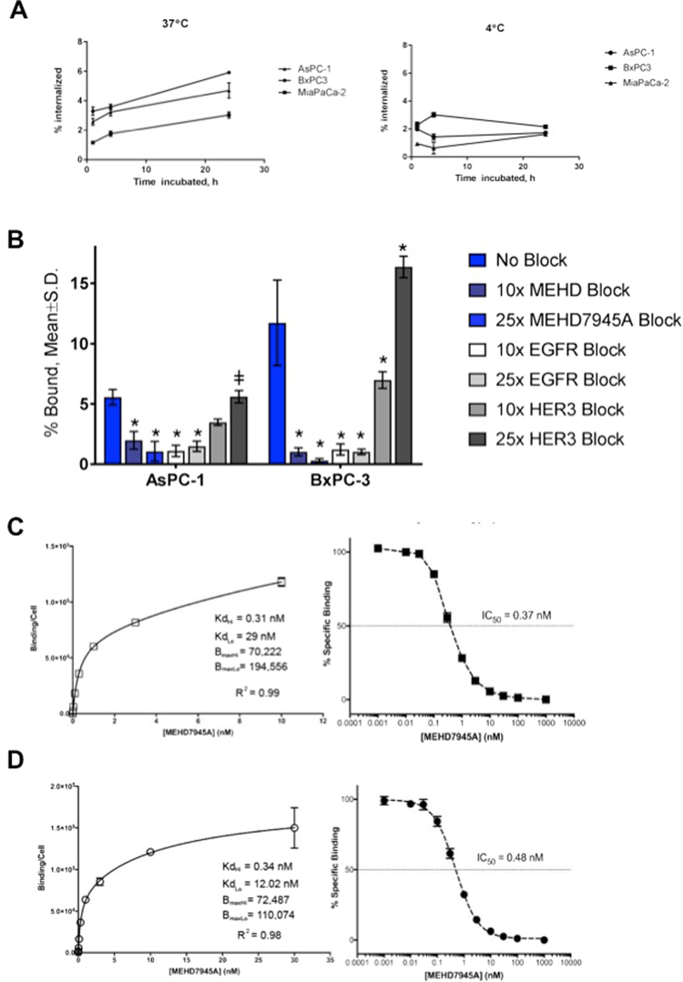
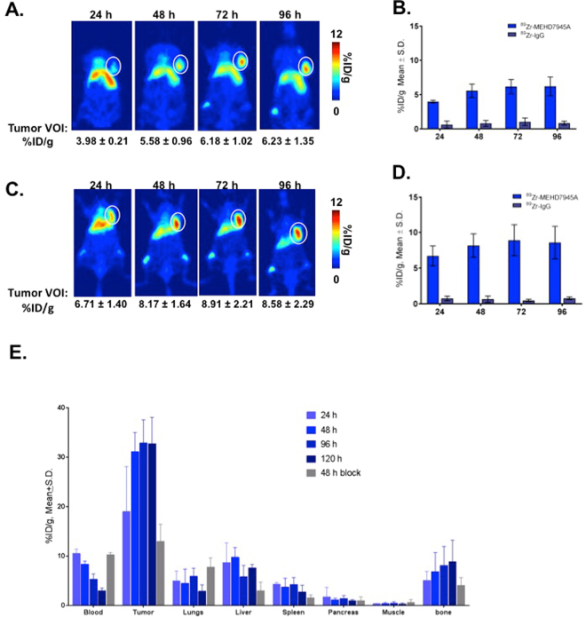
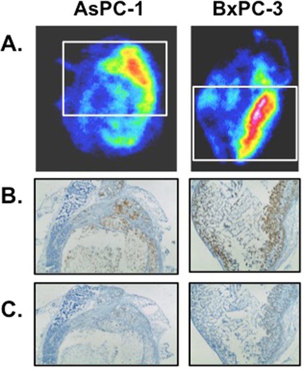
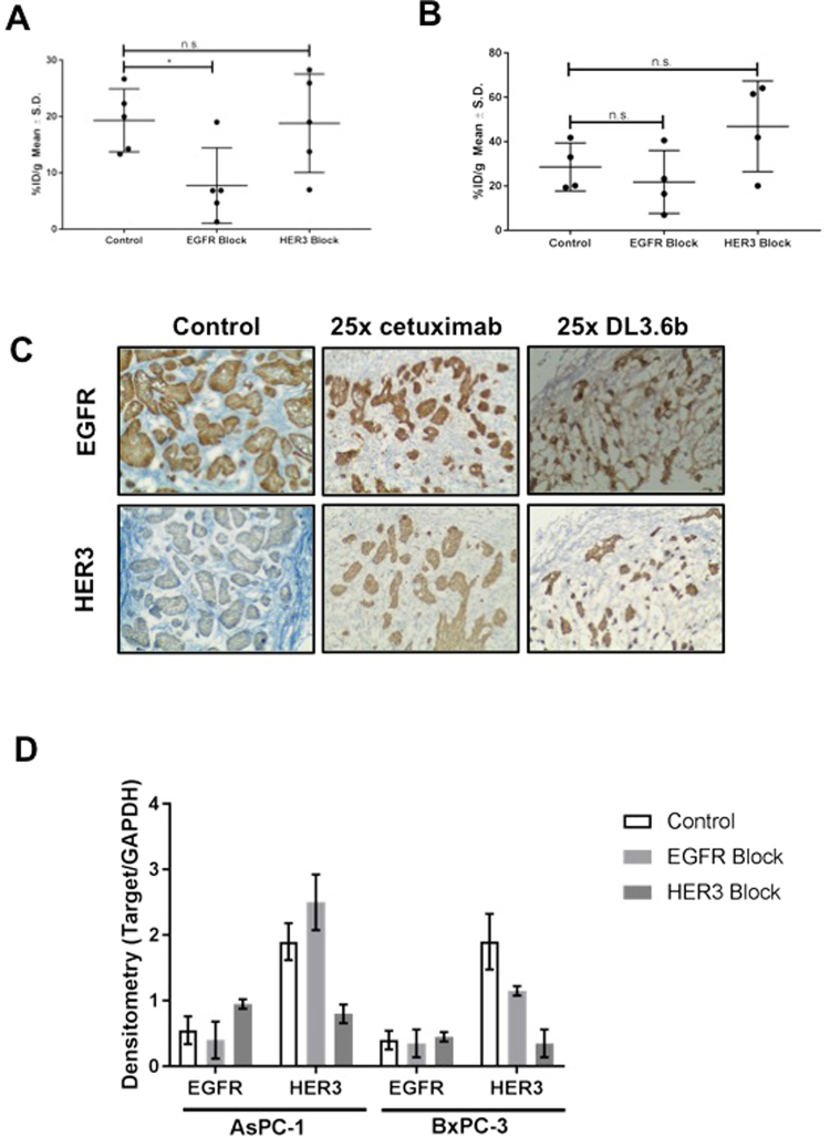
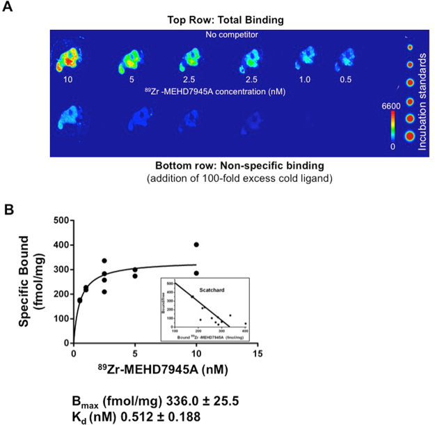
Tumor resistance to treatment paved the way toward the development of single agent drugs that target multiple molecular signatures amplified within the malignancy. The discovered crosstalk between EGFR and HER3 as well as the role of HER3 in mediating EGFR resistance made these two receptor tyrosine kinases attractive targets. MEHD7945A or duligotuzumab is a single immunotherapy agent that dually targets both molecular signatures. In this study, a positron emission tomography (PET) companion diagnostic to MEHD7945A is reported and evaluated in pancreatic cancer. Tumor accretion and whole body pharmacokinetics of 89Zr-MEHD7945A were established. Specificity of the probe for EGFR and/or HER3 was further examined.


Conflict of interest statement

The authors declare no competing interests.

Figures

Figure 1
Figure 1
In vitro characterization. 89Zr-MEHD7945A demonstrated higher internalization rates in BxPC-3 and AsPC-1 at 37 °C (left) compared to the EGFR/HER3-negative Mia PACA2 pancreatic cancer cell line. 89Zr-MEHD7945A showed a decrease in internalization at 4 °C (right) in all cell lines. (A) 89Zr-MEHD7945A demonstrated successful blocking with cold MEHD7945A and cetuximab at 10× and 25× doses. Blocking with DL3.6b at 10× lowered the uptake of the probe; binding was sustained at 25× dose of the anti-HER3 mAb. In both AsPC-1 and BxPC-3. (B) Non-linear regression analysis determined two sets of KD and Bmax for AsPC-1 with an. (C) The KD and Bmax values for BxPC-3 (D) are within the same range as the established values in AsPC-1 with an IC50 ~ 0.37 nM. (*Denote p < 0.01, ǂdenote p < 0.05, compared to no block).
Figure 2
Figure 2
In vivo PET imaging. In AsPC-1 xenografts, tumor volumes-of-interest (VOI) expressed as % ID/g generated from the 89Zr-MEHD7945A PET scans exhibited uptake as early as 24 h p.i., peaking at 72 h p.i. and retained to as long as 96 h p.i. (A) 89Zr-IgG control PET scans showed minimal uptake within the tumor at all time points. (B) Similarly in BxPC-3 tumors, PET scans exhibited uptake at 24 h p.i. with a peak at 72 h p.i. (C) Non-specific tumor uptake using 89Zr-IgG in BxPC-3 xenografts showed nominal accumulation across all time points. (D) Whole body tissue distribution revealed high tumor tissue uptake of the tracer at 24 h p.i,, which plateaued at 48 h through 120 h p.i. in BxPC-3 xenografts. A competitive blocking study using unmodified MEHD7945A at 48 h p.i. displayed at least a two-fold decrease in tumor binding, indicative of the probe’s specificity. (E) Of note, normal pancreas demonstrated minimal non-specific binding on all time points, suggesting that an excellent signal-to-noise contrast can be achieved.
Figure 3
Figure 3
Autoradiography and histology. Autoradiographs (A) of excised AsPC-1 (left) and BxPC-3 (right) tumor sections depicted co-localization of the tracer in areas where EGFR (BxPC-3 3+, AsPC-1 2+) (B) and HER3 (BxPC-3 1+, AsPC-1 2+) (C) are expressed.
Figure 4
Figure 4
In vivo competitive inhibition. In AsPC-1 xenografts, blocking with cetuximab (EGFR block) showed an almost 2-fold decrease in 89Zr-MEHD7945A uptake, whereas blocking HER3 with DL3.6b did not change probe uptake. (A) In BxPC-3 xenografts, blocking with cetuximab (EGFR block) showed a slight decrease in 89Zr-MEHD7945A, whereas blocking with DL3.6b (HER3 block) showed a statistically significant, increase in probe accumulation. (B) IHC staining in BxPC-3 tumors blocked with 25× cetuximab (25x EGFR, left), 25x DL3.6b (25x HER3, middle) or left unblocked (right) were assessed by IHC for EGFR (top) and HER3 (bottom) expression, and showed an increase in EGFR and HER3 in both blocked cohorts. (C) Tumor sections depicted for IHC are shown in 100×. Densitometry analysis of western blots on tumor lysates (n = 2) from AsPC-1 (left) and BxPC-3 (right) that were untreated, exposed to EGFR-block with cetuximab and a HER3-block with DL3.6b. (D) Densitometry is shown as a ratio of target protein/loading control.
Figure 5
Figure 5
Ex vivo competitive binding assay. A digital autoradiograph of AsPC-1 tumor sections displayed saturable, concentration-dependent binding of 89Zr-MEHD7945A upon addition of 100-fold excess cold MEHD7945A. (A) A non-linear regression analysis and Scatchard plot of 89Zr-MEHD7945A plotted against the amount of bound ligand shows the Bmax ~ 336 fmol/mg and a KD ~ 0.51 nM (B).

References

    1. Chandarlapaty S, et al. AKT inhibition relieves feedback suppression of receptor tyrosine kinase expression and activity. Cancer Cell. 2011;19:58–71. doi: 10.1016/j.ccr.2010.10.031. - DOI - PMC - PubMed
    1. Markman B, Tao JJ, Scaltriti M. PI3K pathway inhibitors: better not left alone. Curr Pharm Des. 2013;19:895–906. doi: 10.2174/138161213804547213. - DOI - PubMed
    1. Chakrabarty A, Sanchez V, Kuba MG, Rinehart C, Arteaga CL. Feedback upregulation of HER3 (ErbB3) expression and activity attenuates antitumor effect of PI3K inhibitors. Proc. Natl. Acad. Sci. 2012;109:2718–2723. doi: 10.1073/pnas.1018001108. - DOI - PMC - PubMed
    1. Barton CM, Hall Pa, Hughes CM, Gullick WJ, Lemoine NR. Transforming growth factor alpha and epidermal growth factor in human pancreatic cancer. J. Pathol. 1991;163:111–6. doi: 10.1002/path.1711630206. - DOI - PubMed
    1. Dancer, J., Takei, H., Ro, J. Y. & Lowery-Nordberg, M. Coexpression of EGFR and HER-2 in pancreatic ductal adenocarcinoma: A comparative study using immunohistochemistry correlated with gene amplification by fluorescencent in situ hybridization. Oncol. Rep. (2007). - PubMed

Publication types

MeSH terms