. 2018 Jun 13;8(1):9014.
doi: 10.1038/s41598-018-27336-x.

Avian tail ontogeny, pygostyle formation, and interpretation of juvenile Mesozoic specimens

Affiliations

Avian tail ontogeny, pygostyle formation, and interpretation of juvenile Mesozoic specimens

Dana J Rashid et al. Sci Rep. .

Abstract

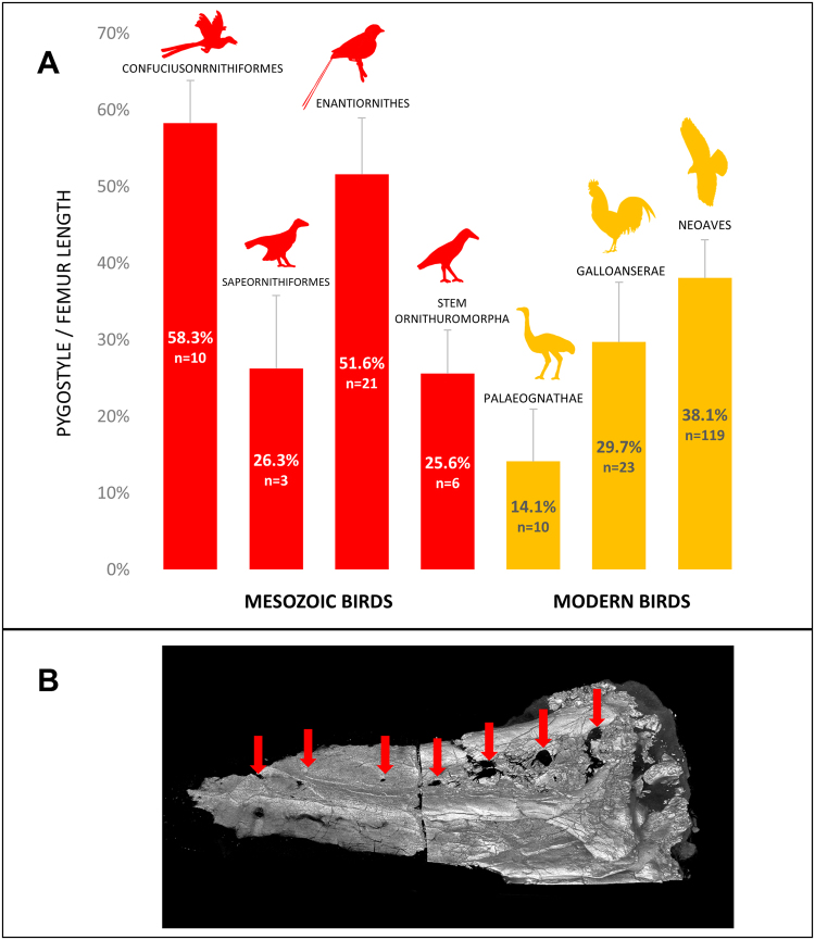
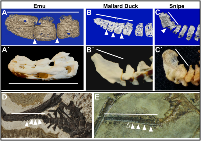
The avian tail played a critical role in the evolutionary transition from long- to short-tailed birds, yet its ontogeny in extant birds has largely been ignored. This deficit has hampered efforts to effectively identify intermediate species during the Mesozoic transition to short tails. Here we show that fusion of distal vertebrae into the pygostyle structure does not occur in extant birds until near skeletal maturity, and mineralization of vertebral processes also occurs long after hatching. Evidence for post-hatching pygostyle formation is also demonstrated in two Cretaceous specimens, a juvenile enantiornithine and a subadult basal ornithuromorph. These findings call for reinterpretations of Zhongornis haoae, a Cretaceous bird hypothesized to be an intermediate in the long- to short-tailed bird transition, and of the recently discovered coelurosaur tail embedded in amber. Zhongornis, as a juvenile, may not yet have formed a pygostyle, and the amber-embedded tail specimen is reinterpreted as possibly avian. Analyses of relative pygostyle lengths in extant and Cretaceous birds suggests the number of vertebrae incorporated into the pygostyle has varied considerably, further complicating the interpretation of potential transitional species. In addition, this analysis of avian tail development reveals the generation and loss of intervertebral discs in the pygostyle, vertebral bodies derived from different kinds of cartilage, and alternative modes of caudal vertebral process morphogenesis in birds. These findings demonstrate that avian tail ontogeny is a crucial parameter specifically for the interpretation of Mesozoic specimens, and generally for insights into vertebrae formation.


Conflict of interest statement

The authors declare no competing interests.

Figures

Figure 1
Figure 1
Timeline of pygostyle formation in the chicken. (A) Alcian blue and picrosirius red histology staining on semi-thin paraffin sections of progressively older pygostyles, mid-sagittal views, distal to the left and dorsal to the top. At 8 days post hatching, D8, intervertebral discs are present and no fusion is observed, but by D168, all ossified pygostyle vertebrae have fused. Intervertebral discs are marked with color-coded arrowheads to track their progressive dismantling by tissue remodeling; the hatched arrowhead indicates a disc in the process of disassemblage during vertebral fusion. The yellow arrow in this and subsequent panels indicates the spinal cord channel. Fusion of chicken pygostyle vertebrae therefore requires 5 months for completion, and occurs in the distal to proximal direction. (B,B′) MicroCT scan of 7–8 week old juvenile chicken pygostyle, mid-sagittal and surface views, respectively; distal to the left. MicroCT scanning confirms unfused pygostyle vertebrae in the juvenile. (C,C′) MicroCT scan of 1.5 year old adult chicken pygostyle, mid-sagittal and surface views, respectively. After pygostyle vertebrae fusion, the spinal cord channel is retained but trabecular bone remodeling removes all traces of intervertebral discs.
Figure 2
Figure 2
Mineralized shapes of tail vertebrae change significantly during ontogeny. (A) Chicken embryonic day E19 wholemount alcian blue and alizarin red; dorsal view, proximal to the top. Alcian blue stains unmineralized cartilage blue and alizarin red stains mineralized tissue red (appears black in this image). In all panels where present, black arrowheads indicate parapophyses. (B) Chicken E19, sagittal view, ossification centers in the pygostyle region; alcian blue and picrosirius red; distal to the left; white arrows indicate ossification centers. (C) Chicken D4, wholemount alcian blue and alizarin red; dorsal view; distal to the left. (D) Chicken 7–8 week old juvenile, pygostyle transverse section, Masson’s Trichrome; dorsal to the top, ventral to the bottom; black arrow indicates spinal cord channel; asterisks indicate unfused medial arch halves. (E) Higher magnification of left parapophysis from (D). (F) Chicken 7–8 week old juvenile, transverse section of left parapophysis, alcian blue and picrosirius red. Compare the red, more sparsely cellular cartilage in this parapophysis tissue with the blue hyaline cartilage in (B) (same stain), indicating disparate matrix staining likely due to increased collagen in the parapophysis. (G) Chicken 7–8 week juvenile, pygostyle transverse fresh cut section, unstained; white arrowheads indicate transverse process extensions. (H) Same tissue as in (G) von Kossa, which stains mineralized tissue black. (I) Chicken 4+ years old, pygostyle transverse section, alcian blue and picrosirius red. Note that trabecular bone remodeling has extended to the tips of transverse processes and parapophyses. (J) MicroCT scan, juvenile Eurypyga helias (LACM 104451) partial tail with pygostyle, dorsal view. (K) MicroCT scan, adult E. helias (LACM 90009) partial tail with pygostyle. Note the mineralized extensions of parapophyses in the adult relative to the juvenile tail vertebrae. Abbreviations: c - centrum, ID - intervertebral disc; NA - neural arch; OF - ossification front; V - vertebra. LACM is the abbreviation for the Los Angeles County Museum of Natural History, followed by the specimen number.
Figure 3
Figure 3
Commonalities and differences in avian caudal vertebral morphogenesis. (A–F) Morphogenesis of parapophyses (Pp) and transverse processes (Tp) occurs by epiphyseal-plate-mediated ossification, but further extension can be achieved by fusion of additional ossified elements. (A) 4.5 month old emu transverse process from a free caudal vertebra, transverse paraffin section, stained with alcian blue and picrosirius red. The morphology of distal transverse process cartilage in the emu is indistinguishable from equivalent staining in chicken parapophyses (see Fig. 2F), but these cartilages differ from the embryonic hyaline cartilage model (Fig. 2B); (B) chicken D126 proximal pygostyle transverse section, von Kossa stained, area in black rectangle magnified in (C) showing nearly complete extension of ossified transverse process by epiphyseal plate-mediated ossification that progressed in the medial to distal direction. (D) Juvenile lesser nighthawk (LACM 111218) microCT dorsal surface view, showing separate ossified elements (yellow arrowheads), analogous to ribs, that fuse into the parapophyses (white arrow) of free caudal vertebrae (pygostyle region noted by white bar). Complete fusion of these bony elements to vertebral bodies can be seen in (E) a microCT dorsal view of an adult lesser nighthawk (LACM 73857). (F–H) MicroCT dorsal views of juvenile tails with no evidence of caudal ribs; (F) juvenile red-headed woodpecker (LACM 111600), this specimen is at a similar ontogenetic stage, but shows no evidence of the rib-like bony elements. Caudal ribs are also absent in (G) a juvenile western screech-owl (LACM 111461) and in (H) a developmentally more advanced elegant tern (LACM 116139).
Figure 4
Figure 4
A ventral furrow in caudal vertebrae is a common extant avian trait. A ventral furrow or concave surface in extant avians shelters the ventral tail vasculature. This vasculature includes the branched aorta artery, as well as caudal veins. (A) A transverse paraffin section of a 7–8 week old chicken juvenile pygostyle stained with alcian blue and picrosirius red; blood vessels are noted by the black arrowheads. A ventral caudal furrow is evident in the proximal caudal vertebrae in (B) a red-tailed hawk, Buteo jamaicensis skeletal tail (MOR 1230), especially in the pygostyle region, and in the entire tail in (C) a golden eagle, Aquila chrysaetos (MOR 116) (ventral views). (D) Enlarged ventrolateral view of the golden eagle pygostyle, showing the deeply recessed furrow. MOR is the abbreviation for the Museum of the Rockies.
Figure 5
Figure 5
Some Mesozoic birds formed a relatively longer pygostyle than extant birds. (A) The percent of adult pygostyle length to femur length was plotted for four different Cretaceous (red) and three different extant (orange) bird groups. The data shows great variation in relative pygostyle lengths overall, and suggests that confuciusornithiformes and enantiornithes incorporated more vertebrae into their pygostyle (n: number of specimens measured). (B) MicroCT scan of a Confuciusornis pygostyle (NGMC 98-8-2); distal to the left, dorsal to the top. Red arrows indicate intervertebral foramen from incompletely fused neural arches which theoretically correlate to the original number of pygostyle vertebrae before fusion. The break in the middle is due to a thin bone section removed for histology, with the two pygostyle halves digitally reconstructed. Silhouettes were either taken from public domain images on phylopic.org (Protopteryx, Matt Martyniuk; Gallus, Steven Traver; and Buteo, Lauren Anderson), or drawn by D. Rashid (Confuciusornithiformes, Sapeornithiformes, Stem Ornithuromorpha, and Palaeognathae silhouettes). NGMC is the abbreviation for the National Geological Museum of China.
Figure 6
Figure 6
Unfused juvenile pygostyles in extant and Cretaceous birds. White arrows indicate intervertebral spaces (empty arrowhead indicates possible intervertebral space); white bars indicate linear extent of pygostyles; all pygostyles distal to the left, dorsal to the top. (A) 5.5 month old emu pygostyle, microCT mid-sagittal view. (A′) Adult emu pygostyle. (B) Juvenile mallard duck distal tail, microCT mid-sagittal view. (B′) Adult mallard duck distal tail. (C) Juvenile Wilson’s snipe distal tail, microCT sagittal view. (C′) Adult Wilson’s snipe distal tail. (D) Juvenile Archaeorhynchus IVPP 17075. (E) Juvenile enantiornithine, IVPP 15664.

Similar articles

Cited by

References

    1. Chatterjee, S. The Rise of Birds: 225 Million Years of Evolution, (Johns Hopkins University Press) (1997).
    1. Chiappe, L. M. & Witmer, L. M. Mesozoic Birds: Above the Heads of Dinosaurs, (Berkeley, Los Angeles, and London: University of California Press) (2002).
    1. Heers AM, Dial KP. From extant to extinct: locomotor ontogeny and the evolution of avian flight. Trends Ecol Evol. 2012;27:296–305. doi: 10.1016/j.tree.2011.12.003. - DOI - PubMed
    1. Huxley TH. On the animals which are most nearly intermediate between birds and reptiles. Geol. Mag. 1868;5:357–365. doi: 10.1017/S0016756800207784. - DOI
    1. Huxley TH. Further Evidence of the Affinity between the Dinosaurian Reptiles and Birds. Quarterly Journal of the Geological Society. 1870;26:12–31. doi: 10.1144/GSL.JGS.1870.026.01-02.08. - DOI

Publication types

LinkOut - more resources