

The fibrogenic chemokine CCL18 is associated with disease severity in Erdheim-Chester disease
- PMID: 29900045
- PMCID: PMC5993512
- DOI: 10.1080/2162402X.2018.1440929
The fibrogenic chemokine CCL18 is associated with disease severity in Erdheim-Chester disease
Abstract
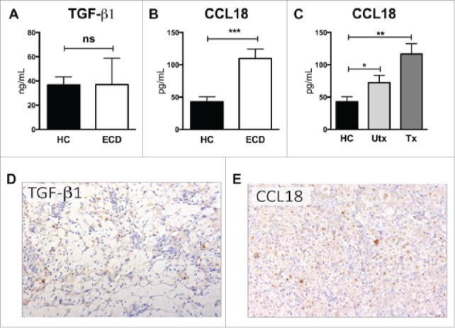
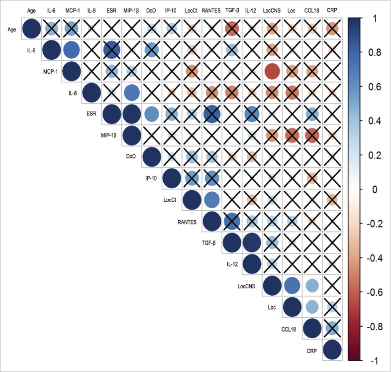
Erdheim-Chester disease (ECD) is a rare histiocytosis, characterized by xanthogranulomatous tissue infiltration by foamy histiocytes. Fibrosis, a histologic hallmark of ECD, is responsible for lesion growth and clinical manifestations. Unraveling molecular fibrotic pathway in ECD would allow the identification of new pharmacologic targets. In this study, we evaluated serum and tissue samples from a large cohort of ECD patients focusing on two major pro-fibrotic mediators, TGF-β1 and chemokine ligand 18 (CCL18). We found a marked increase in CCL18 but not TGF-β1 levels in serum and lesions of ECD patients (p < 0.001), independently of treatment status and consistently over time. Using a linear mathematical model, we also found that elevated CCL18 serum levels correlate with both number and severity of disease localizations. These findings suggest the involvement of CCL18-induced fibrosis in ECD pathogenesis, providing a rationale for exploring CCL18 inhibition as a treatment for progressive fibrosis in ECD.
Keywords: CCL-18; Erdheim-Chester disease; fibrosis.
Figures
References
-
- Campochiaro C, Tomelleri A, Cavalli G, Berti A, Dagna L. Erdheim-Chester disease. Eur J Intern Med [Internet]. 2015;26:223–9. Available from: http://linkinghub.elsevier.com/retrieve/pii/S0953620515000771. doi: 10.1016/j.ejim.2015.03.004. - DOI - PubMed
-
- Cavalli G, Guglielmi B, Berti A, Campochiaro C, Sabbadini MG, Dagna L. The multifaceted clinical presentations and manifestations of Erdheim-Chester disease: comprehensive review of the literature and of 10 new cases. Ann Rheum Dis [Internet]. 2013;72:1691–5. Available from: http://www.ncbi.nlm.nih.gov/pubmed/23396641. doi: 10.1136/annrheumdis-2012-202542. - DOI - PubMed
-
- Cavalli G, Berti A, Campochiaro C, Dagna L. Diagnosing Erdheim–Chester disease. Ann Rheum Dis [Internet]. 2013;72:e19 LP-e19 Available from: http://ard.bmj.com/content/72/7/e19.abstract - PubMed
-
- Haroche J, Charlotte F, Arnaud L, Deimling A Von, Hélias-rodzewicz Z, Hervier B, Cohen-aubart F, Launay D, Lesot A, Mokhtari K, et al.. High prevalence of BRAF V600E mutations in Erdheim-Chester disease but not in other non-Langerhans cell histiocytoses Brief report High prevalence of BRAF V600E mutations in Erdheim-Chester disease but not in other non-Langerhans cell histiocytoses. Blood 2012;120:2700–3. - PubMed
-
- Cangi MG, Biavasco R, Cavalli G, Grassini G, Dal-Cin E, Campochiaro C, Guglielmi B, Berti A, Lampasona V, von Deimling A, et al.. BRAFV600E-mutation is invariably present and associated to oncogene-induced senescence in Erdheim-Chester disease. Ann Rheum Dis [Internet]. 2015;74:1–7. Available from: http://www.ncbi.nlm.nih.gov/pubmed/24671772 - PubMed
Publication types
LinkOut - more resources
Full Text Sources
Other Literature Sources