. 2018 May 30:5:164.
doi: 10.3389/fmed.2018.00164. eCollection 2018.

Fluorescence Exclusion: A Simple Method to Assess Projected Surface, Volume and Morphology of Red Blood Cells Stored in Blood Bank

Affiliations

Fluorescence Exclusion: A Simple Method to Assess Projected Surface, Volume and Morphology of Red Blood Cells Stored in Blood Bank

Camille Roussel et al. Front Med (Lausanne). .

Abstract

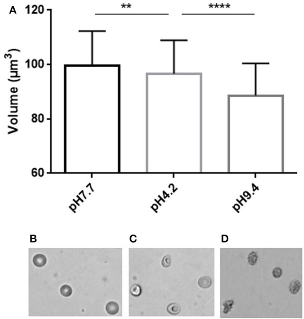
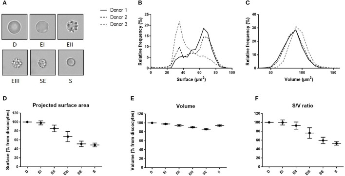
Red blood cells (RBC) ability to circulate is closely related to their surface area-to-volume ratio. A decrease in this ratio induces a decrease in RBC deformability that can lead to their retention and elimination in the spleen. We recently showed that a subpopulation of "small RBC" with reduced projected surface area accumulated upon storage in blood bank concentrates, but data on the volume of these altered RBC are lacking. So far, single cell measurement of RBC volume has remained a challenging task achieved by a few sophisticated methods some being subject to potential artifacts. We aimed to develop a reproducible and ergonomic method to assess simultaneously RBC volume and morphology at the single cell level. We adapted the fluorescence exclusion measurement of volume in nucleated cells to the measurement of RBC volume. This method requires no pre-treatment of the cell and can be performed in physiological or experimental buffer. In addition to RBC volume assessment, brightfield images enabling a precise definition of the morphology and the measurement of projected surface area can be generated simultaneously. We first verified that fluorescence exclusion is precise, reproducible and can quantify volume modifications following morphological changes induced by heating or incubation in non-physiological medium. We then used the method to characterize RBC stored for 42 days in SAG-M in blood bank conditions. Simultaneous determination of the volume, projected surface area and morphology allowed to evaluate the surface area-to-volume ratio of individual RBC upon storage. We observed a similar surface area-to-volume ratio in discocytes (D) and echinocytes I (EI), which decreased in EII (7%) and EIII (24%), sphero-echinocytes (SE; 41%) and spherocytes (S; 47%). If RBC dimensions determine indeed the ability of RBC to cross the spleen, these modifications are expected to induce the rapid splenic entrapment of the most morphologically altered RBC (EIII, SE, and S) and further support the hypothesis of a rapid clearance of the "small RBC" subpopulation by the spleen following transfusion.

Keywords: fluorescence exclusion; red blood cell morphology; red blood cell storage; red blood cell volume; red blood cells; transfusion.


Figures

Figure 1
Figure 1
(A) Top: Principle of the fluorescence exclusion method, bottom: design of the microfluidic chamber. (B,C) Brightfield and fluorescence images of the same field (x20) showing RBC stored for 42 days in SAG-M (healthy donor) scale bar = 20 μm.
Figure 2
Figure 2
Stepwise preparation of chips: (A) Chips were made using a mixture 1:10 of PDMS (polydimethylsiloxane) and its cross linker (Sylgard 184) (B) The mixture was degased to remove air bubbles, (C) poured into the mold and (D) cured at 66°C for 2 h. (E) After demolding, inlets and outlets were created with 2 or 3 mm punchers and (F) the surface was cleaned using isopropanol, air gun and tape before bonding. (G) Chambers were bond on glass-bottomed petri dishes (Fluorodish) using air plasma cleaner (Harrick) or a corona SB (Elveflow).
Figure 3
Figure 3
Fluorescence exclusion to measure RBC volume: a precise and reproducible method. (A) Normalized frequency histogram of RBC volume distribution from a healthy donor at day 10 of storage in SAG-M. (B) Individual volume of RBC from a healthy donor measured using 2 differents chips manufactured from the same mold (chip A and chip B). (C) Fluorescence (left) and brightfield (right) images (20x) of control RBC (top) or 100% heated RBC (bottom) scale bar = 20 μm; (D) Normalized frequency histogram of RBC volume distribution, and (E) Mean volume (+/−SEM) of samples containing increasing concentrations of heated-RBC.
Figure 4
Figure 4
Simultaneous determination of RBC volume and morphology using fluorescence exclusion and microscopy. (A) Mean (standard deviation) volume of RBC samples exposed to physiological (left), low (center) and high (right) pH. Stars represent the significance level of the Mann-Whitney statistic test, ** when p < 0.01, **** when p < 0.0001 (B–D) Representative brightfield images of RBC in the same samples.
Figure 5
Figure 5
Fluorescence exclusion shows a decrease in surface to volume ratio during storage of red blood cell concentrates. (A) Morphological categories of RBC as defined in the Material and Methods section, namely discocytes (D), echinocytes I (EI), echinocytes II (EII), echinocytes III (EIII), spheroechinocytes (SE) and spherocytes (S). (B–C) Normalized frequency of RBC projected surface area (μm2) and RBC volume (μm3) of RBC concentrates after 42 days of storage in SAG-M (n = 3). (D-F) Mean (SD) projected surface area, volume and projected surface area-to-volume ratio normalized to the value of discocytes (D).

References

    1. Mohandas N, Clark MR, Jacobs MS, Shohet SB. Analysis of factors regulating erythrocyte deformability. J Clin Invest. (1980) 66:563–73. 10.1172/JCI109888 - DOI - PMC - PubMed
    1. Waugh RE, Narla M, Jackson CW, Mueller TJ, Suzuki T, Dale GL. Rheologic properties of senescent erythrocytes: loss of surface area and volume with red blood cell age. Blood (1992) 79:1351–8. - PubMed
    1. Waugh RE, Sarelius IH. Effects of lost surface area on red blood cells and red blood cell survival in mice. Am J Physiol. (1996) 271(6 Pt 1):C1847–1852. 10.1152/ajpcell.1996.271.6.C1847 - DOI - PubMed
    1. Perrotta S, Gallagher PG, Mohandas N. Hereditary spherocytosis. Lancet Lond Engl. (2008) 372:1411–26. 10.1016/S0140-6736(08)61588-3 - DOI - PubMed
    1. Gallagher PG. Disorders of red cell volume regulation. Curr Opin Hematol. (2013) 20:201–7. 10.1097/MOH.0b013e32835f6870 - DOI - PubMed

LinkOut - more resources