Review
. 2018 Oct;19(10):761-769.
doi: 10.1111/tra.12587. Epub 2018 Jul 17.

Mechanisms of lysosomal positioning and movement

Affiliations
Review

Mechanisms of lysosomal positioning and movement

Birol Cabukusta et al. Traffic. 2018 Oct.

Abstract

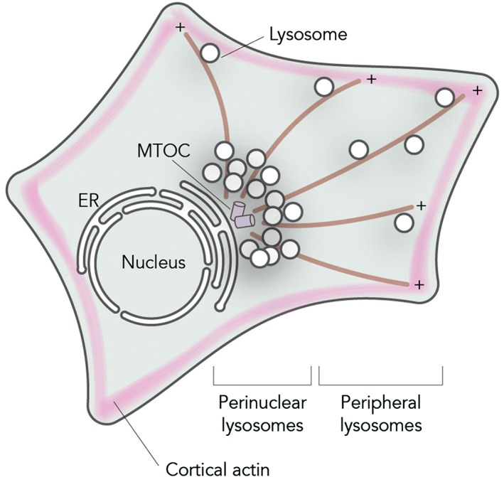
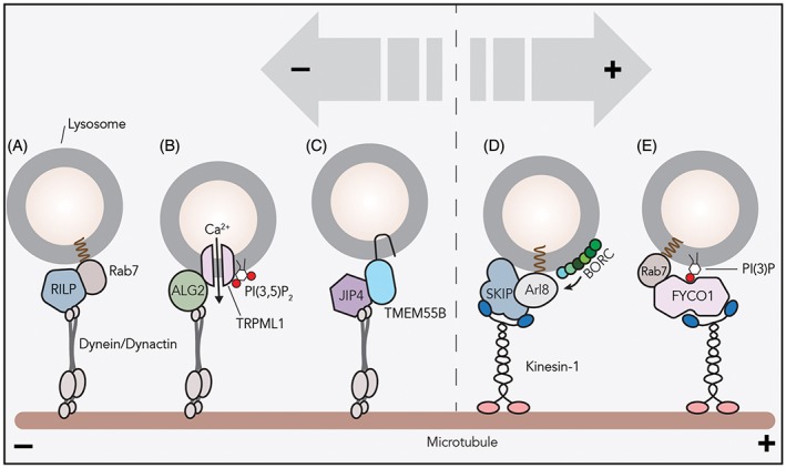
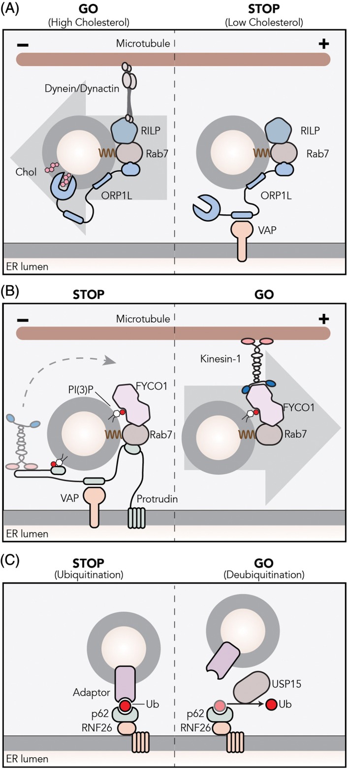
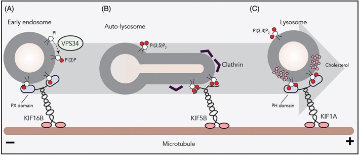
Lysosomes are highly dynamic organelles that can move rapidly throughout the cell. They distribute in a rather immobile pool located around the microtubule-organizing center in a "cloud," and a highly dynamic pool in the cell periphery. Their spatiotemporal characteristics allow them to carry out multiple biological functions, such as cargo degradation, antigen presentation and plasma membrane repair. Therefore, it is not surprising that lysosomal dysfunction underlies various diseases, including cancer, neurodegenerative and autoimmune diseases. In most of these biological events, the involvement of lysosomes is dependent on their ability to move throughout the cytoplasm, to find and fuse to the correct compartments to receive and deliver substrates for further handling. These dynamics are orchestrated by motor proteins moving along cytoskeletal components. The complexity of the mechanisms responsible for controlling lysosomal transport has recently been appreciated and has yielded novel insights into interorganellar communication, as well as lipid-protein interplay. In this review, we discuss the current understanding of the mechanisms of lysosomal transport and the molecular machineries that control this mobility.

Keywords: Arl GTPases; ORP1L; RNF26; Rab GTPases; TOLLIP; cholesterol; dendritic cells; dynein; endoplasmic reticulum; kinesins; late endosome; lysosome; mTOR; membrane contact sites; myosins; phosphatidylinositol phosphate.


Figures

Figure 1
Figure 1
Intracellular distribution of lysosomes. Lysosomes can be found in two intracellular locations: a relatively immobile pool of perinuclear lysosomes clustered around the MTOC and a set of peripheral lysosomes moving fast along microtubules in a stop‐and‐go manner or are found attached to peripheral actin networks
Figure 2
Figure 2
Many mechanisms of lysosomal transport. Various mechanisms of lysosomal recruitment of motor proteins are reported. A, The small GTPase Rab7 recruits effector RILP and the dynein/dynactin motor to lysosomes for minus‐end transport. B, Calcium plays a crucial role in lysosomal positioning. PI(3,5)P2 on lysosomal‐delimiting membranes activates the TRPML1 channel to stimulate calcium efflux. Then, the cytosolic calcium sensor ALG2 recruits the dynein/dynactin complex to TRPML1‐containing lysosomes. C, Transmembrane protein TMEM55B interacts with the dynein adaptor JIP4 and facilitates minus‐end transport. TMEM55B levels are controlled transcriptionally: depletion of nutrients or cholesterol upregulates TMEM55B transcription via autophagy‐associated transcription factors. D, BORC, a multisubunit complex on the lysosomal membrane, recruits Arl8b to lysosomes. The small GTPase Arl8b interacts with effector SKIP for lysosomal localization of kinesin‐1. E, The kinesin adaptor FYCO1 interacts with active Rab7 and PI(3)P on lysosomal membranes to recruit kinesin‐1 to lysosomes
Figure 3
Figure 3
ER‐lysosome membrane contact sites control lysosomal transport and positioning. A, The Rab7 effector ORP1L localizes to lysosomes by interacting with Rab7 and phosphoinositides on the lysosomal membrane. When cholesterol is sufficient on the lysosomal‐ or autophagosomal‐delimiting membrane, ORP1L found in a closed conformation that mediates dynactin/dynein complex assembly and minus‐end transport via RILP. Under low‐cholesterol conditions, ORP1L adopts an open conformation and exposes an FFAT motif that allows interaction with the ER protein VAP. The resulting membrane contact site forces the release of the dynein/dynactin complex. B, ER‐bound protrudin interacts with the late Rab7 and PI(3)P and loads kinesin‐1 to Rab7‐effector FYCO1 on PI(3)P‐containing lysosomes. C, The ubiquitination of p62/SQSTM1 adaptor by the ER‐localized ubiquitin ligase RNF26 is recognized by TOLLIP that has a ubiquitin‐binding domain. This complex dictates perinuclear positioning of lysosomes. The cytosolic deubiquitinating enzyme USP15 mediates the release of lysosomes
Figure 4
Figure 4
The role of phosphoinositides on lysosomal transport. A, PI(3)P, a phosphoinositide marking the endosomal system, is generated on the early endosomal membrane by the PI kinase VPS34. PI(3)P on early endosome serves as a docking station for KIF16B (kinesin‐3) to drive plus‐end transport. B, Clathrin on autophagosomal membranes causes clustering of PI(4,5)P2 that allows the binding of the KIF5B (kinesin‐1) onto the autophagosome membrane. KIF5B‐driven plus‐end transport results in autophagosome tubulation and proto‐lysosome formation. C, Formation of membrane microdomains by cholesterol and sphingomyelin or by glycosphingolipids on lysosomal membranes facilitates the interaction between the PH domain of KIF1A (kinesin‐3) and PI(4,5)P2

References

    1. Bakker J, Spits M, Neefjes J, Berlin I. The EGFR odyssey – from activation to destruction in space and time. J Cell Sci. 2017;130(24):4087‐4096. 10.1242/jcs.209197. - DOI - PubMed
    1. Neefjes J, Jongsma MML, Berlin I. Stop or go? Endosome positioning in the establishment of compartment architecture, dynamics, and function. Trends Cell Biol. 2017;27(8):580‐594. 10.1016/j.tcb.2017.03.002. - DOI - PubMed
    1. Jongsma MLLM, Berlin I, Wijdeven RHHM, et al. An ER‐associated pathway defines endosomal architecture for controlled cargo transport. Cell. 2016;166(1):152‐166. 10.1016/j.cell.2016.05.078. - DOI - PMC - PubMed
    1. Johnson DE, Ostrowski P, Jaumouillé V, Grinstein S. The position of lysosomes within the cell determines their luminal pH. J Cell Biol. 2016;212(6):677‐692. 10.1083/jcb.201507112. - DOI - PMC - PubMed
    1. Encarnação M, Espada L, Escrevente C, et al. A Rab3a‐dependent complex essential for lysosome positioning and plasma membrane repair. J Cell Biol. 2016;213(6):631‐640. 10.1083/jcb.201511093. - DOI - PMC - PubMed

Publication types