. 2018 Sep;42(3):1247-1256.
doi: 10.3892/ijmm.2018.3716. Epub 2018 Jun 5.

CDMP1 overexpression mediates inflammatory cytokine‑induced apoptosis via inhibiting the Wnt/β‑Catenin pathway in rat dorsal root ganglia neurons

Affiliations

CDMP1 overexpression mediates inflammatory cytokine‑induced apoptosis via inhibiting the Wnt/β‑Catenin pathway in rat dorsal root ganglia neurons

Zhongwei Jia et al. Int J Mol Med. 2018 Sep.

Abstract

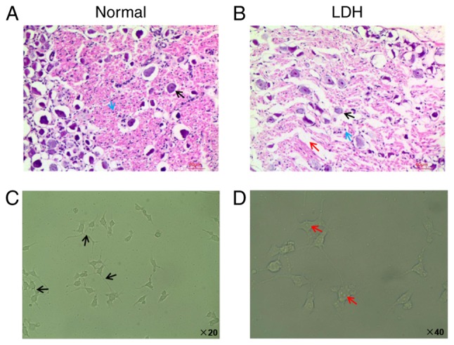
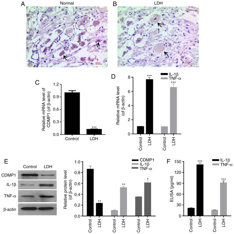
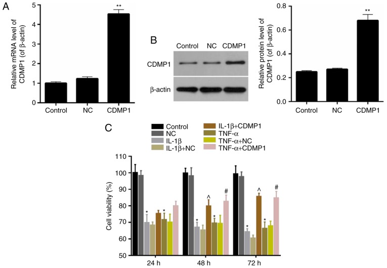
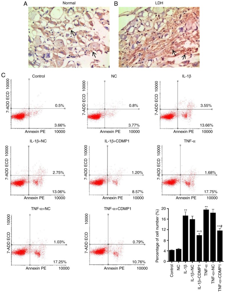
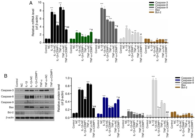
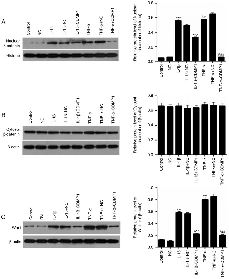
Cartilage‑derived morphogenetic protein‑1 (CDMP1) is a polypeptide growth factor with specific cartilage inducibility, which is predominantly expressed in the developmental long bone cartilage core and in the pre‑cartilage matrix in the embryonic stage. The aim of the present study was to investigate the roles and the mechanisms of CDMP1 overexpression on the apoptosis of rat dorsal root ganglia (DRG) neurons that were induced by inflammatory cytokines. Cell counting Kit‑8 assay, flow cytometry and TdT‑mediated dUTP nick‑end labeling assay were performed to examine cell viability and apoptosis. ELISA, hematoxylin and eosin staining and immunohistochemistry assays were performed to examine the levels of several factors in DRG tissues. Western blot analysis and reverse transcription‑quantitative polymerase chain reaction assays were used to determine the mRNA and protein expression levels, respectively. The results demonstrated that CDMP1 expression was downregulated, while inflammatory cytokine expression was upregulated in DRG tissues derived from lumbar disc herniation (LDH) model rats. In addition, DRG cells from LDH rats exhibited increased apoptosis compared with control rats. CDMP1 overexpression enhanced the cell viability of inflammatory cytokine‑induced DRG cells, and suppressed the apoptosis of inflammatory cytokine‑induced DRG cells via regulating the expression levels of Caspase‑3/8/9, BCL2 apoptosis regulator, and BCL2 associated X. Furthermore, CDMP1 overexpression was demonstrated to affect the Wnt/β‑Catenin pathway in the inflammatory cytokine‑induced DRG cells. In conclusion, the present findings suggested that CDMP1 overexpression mediated inflammatory cytokine‑induced apoptosis via Wnt/β‑Catenin signaling in rat DRG cells.


Figures

Figure 1
Figure 1
Characterization of DRG tissues and cells. Hematoxylin-eosin staining was performed on the DRG tissues from (A) normal rats and (B) LDH model rats. The black arrow indicates the nucleus. The blue arrow indicates the Nissl bodies. The red arrow indicated the intercellular space. Phase-contract images of the DRG cells at magnification, (C) ×20 and (D) ×40 were observed under a fluorescence microscope. The black arrows indicate the cell network structure. The red arrows indicate the clear nucleus. DRG, dorsal root ganglia; LDH, lumbar disc herniation.
Figure 2
Figure 2
CDMP1 expression is downregulated and inflammatory cytokine expression is upregulated in DRG tissues from LDH model rats. (A and B) Immunohistochemistry was performed to evaluate the expression levels of CDMP1 in DRG tissues from (A) normal rats and from (B) LDH model rats. The black arrows indicate CDMP1-positive cells. (C) mRNA levels of CDMP1 in DRG tissues from normal and LDH rats. (D) mRNA levels of IL-1β and TNF-α in DRG tissues from normal and LDH rats. (E) Western blot assay and (F) ELISA results for the protein levels of CDMP1, IL-1β and TNF-α in DRG tissues from normal and LDH rats. *P<0.05, **P<0.01, and ***P<0.001 vs. control. CDMP1, cartilage-derived morphogenetic protein-1; DRG, dorsal root ganglia; LDH, lumbar disc herniation; IL, interleukin; TNF, tumor necrosis factor.
Figure 3
Figure 3
CDMP1 overexpression enhances the cell viability of inflammatory cytokine-induced DRG cells. (A) mRNA and (B) protein levels of CDMP1 in DRG cells transfected with either empty vector (NC) or CDMP1-expressing plasmid (CDMP1). (C) Cell viability of DRG cells in various treatment groups was measured by CCK-8 assay. *P<0.05 and **P<0.01 vs. NC; ^P<0.05 vs. IL-1β+NC; #P<0.05 vs. TNF-α+NC. CDMP1, cartilage-derived morphogenetic protein-1; DRG, dorsal root ganglia; NC, negative control empty vector transfection group; IL, interleukin; TNF, tumor necrosis factor.
Figure 4
Figure 4
CDMP1 overexpression suppresses the apoptosis of inflammatory cytokine-induced DRG cells. (A and B) TUNEL was performed on DRG tissues from normal rats and from LDH model rats in order to examine the apoptosis ratio of DRG cells. The black arrows indicate positive staining. (C) Representative plots and quantification of flow cytometry results. **P<0.01 vs. NC; ^P<0.05 vs. IL-1β+NC; #P<0.05 vs. TNF-α+NC. CDMP1, cartilage-derived morphogenetic protein-1; DRG, dorsal root ganglia; LDH, lumbar disc herniation; NC, negative control empty vector transfection group; IL, interleukin; TNF, tumor necrosis factor.
Figure 5
Figure 5
CDMP1 overexpression regulates the expression of apoptosis-associated proteins in DRG cells. (A) mRNA and (B) protein expression levels of Caspase-3, Caspase-8, Caspase-9, Bax and Bcl-2 were examined in control DRG cells, DRG cells that were transfected with empty vector (NC), and inflammatory cytokine-induced DRG cells that were transfected with either empty vector and CDMP1-expressing vector. *P<0.05, **P<0.01, and ***P<0.001 vs. NC; ^P<0.05 and ^^P<0.01 vs. IL-1β+NC; #P<0.05 and ##P<0.01 vs. TNF-α+NC. CDMP1, cartilage-derived morphogenetic protein-1; DRG, dorsal root ganglia; Bax, BCL2 associated X; Bcl-2, BCL2 apoptosis regulator; NC, negative control empty vector transfection group; IL, interleukin; TNF, tumor necrosis factor.
Figure 6
Figure 6
CDMP1 overexpression represses the Wnt/β-Catenin pathway in inflammatory cytokine-induced DRG cells. Western blot assay was performed on the expression levels of (A) nuclear β-Catenin, (B) cytosolic β-Catenin, and (C) Wnt1. *P<0.05, **P<0.01, and ***P<0.001 vs. NC; ^P<0.05 and ^^P<0.01 vs. IL-1β+NC; ##P<0.01 and ###P<0.001 vs. TNF-α+NC. CDMP1, cartilage-derived morphogenetic protein-1; DRG, dorsal root ganglia; NC, negative control empty vector transfection group; IL, interleukin; TNF, tumor necrosis factor.

Similar articles

Cited by

References

    1. Olmarker K, Larsson K. Tumor necrosis factor alpha and nucleus-pulposus-induced nerve root injury. Spine (Phila Pa 1976) 1998;23:2538–2544. doi: 10.1097/00007632-199812010-00008. - DOI - PubMed
    1. Olmarker K, Rydevik B, Nordborg C. Autologous nucleus pulposus induces neurophysiologic and histologic changes in porcine cauda equina nerve roots. Spine (Phila Pa 1976) 1993;18:1425–1432. doi: 10.1097/00007632-199318110-00005. - DOI - PubMed
    1. Raghavendra V, Tanga F, Rutkowski MD, DeLeo JA. Anti-hyperalgesic and morphine-sparing actions of propentofylline following peripheral nerve injury in rats: Mechanistic implications of spinal glia and proinflammatory cytokines. Pain. 2003;104:655–664. doi: 10.1016/S0304-3959(03)00138-6. - DOI - PubMed
    1. Genevay S, Finckh A, Payer M, Mezin F, Tessitore E, Gabay C, Guerne PA. Elevated levels of tumor necrosis factor-alpha in periradicular fat tissue in patients with radiculopathy from herniated disc. Spine (Phila Pa 1976) 2008;33:2041–2046. doi: 10.1097/BRS.0b013e318183bb86. - DOI - PubMed
    1. Lee S, Moon CS, Sul D, Lee J, Bae M, Hong Y, Lee M, Choi S, Derby R, Kim BJ, et al. Comparison of growth factor and cytokine expression in patients with degenerated disc disease and herniated nucleus pulposus. Clin Biochem. 2009;42:1504–1511. doi: 10.1016/j.clinbiochem.2009.06.017. - DOI - PubMed

Substances