

The roles of the TLR/NF‑κB signaling pathway in the mutual interactions between the lung and the large intestine
- PMID: 29901105
- PMCID: PMC6072181
- DOI: 10.3892/mmr.2018.9111
The roles of the TLR/NF‑κB signaling pathway in the mutual interactions between the lung and the large intestine
Abstract
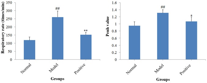
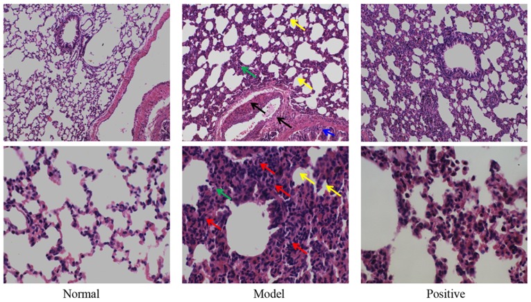
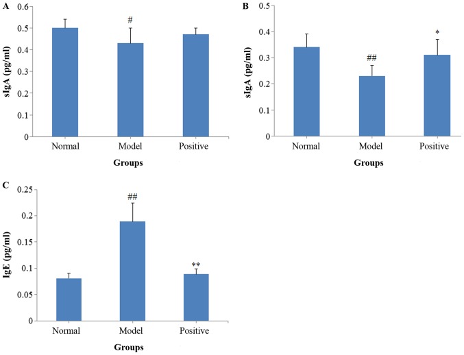
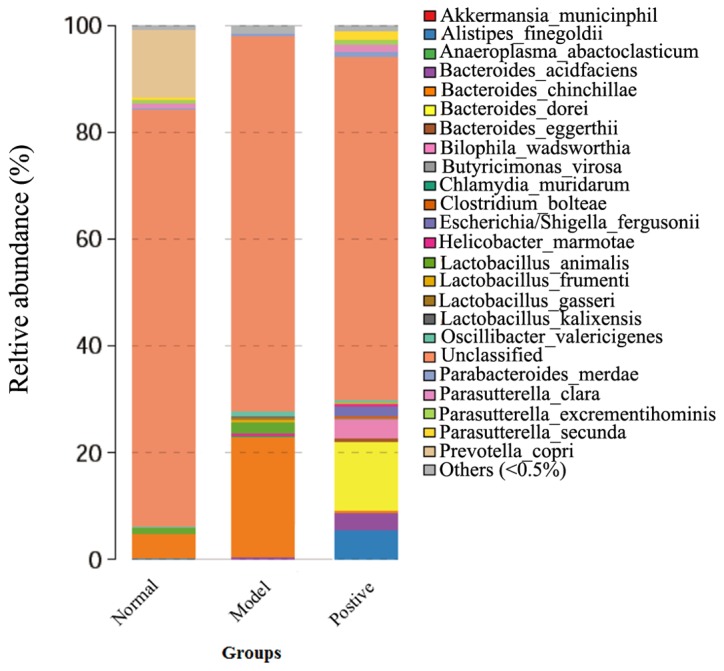
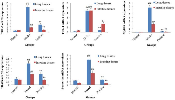
The 'exterior-interior relationship between the lung and the large intestine' is a classical basic theory in Traditional Chinese Medicine. The present study aimed to investigate the roles of the toll like receptor/nuclear factor‑κB (TLR/NF‑κB) signaling pathway in the mutual interactions between the lung and the large intestine. A rat model of allergic asthma complicated with intestinal flora disorder was established by oral administration of Candida albicans and intraperitoneal injection with ovalbumin. The number of inflammatory cells and expression levels immunoglobulin (Ig)E, secretory IgA, interleukin (IL)‑4 and interferon‑γ in serum and bronchoalveolar lavage fluid were subsequently measured. Bacterial colonies and expression of 16S ribosomal DNA were studied in feces samples and pathological alterations of lung tissues were identified. Furthermore, the expression levels of genes associated with the TLR/NF‑κB signaling pathway in the lung and intestinal tissues were determined by reverse transcription‑quantitative polymerase chain reaction. The results of the present study indicated that, in the rat model of allergic asthma complicated with intestinal flora disorder, the expression levels of IL‑4 and IgE, and the numbers of inflammatory cells and C. albicans increased, and marked inflammatory cell infiltration was observed in lung tissues, suggesting that the animal model was successfully established. Furthermore, the present results revealed the mRNA expression levels of genes associated with the TLR/NF‑κB signaling (including myeloid differentiation primary response 88, TNF receptor associated factor 6 and β‑arrestin) were upregulated in both of the lung and intestinal tissues of the model group rats. Collectively, the results demonstrated that the TLR/NF‑κB signaling may serve roles in the mutual interactions between the lung and the large intestine, and TLR and NF‑κB may be potential targets for the treatment of lung diseases complicated with intestinal disorders.
Figures

Similar articles
-
[Role of immune deviation by toll-liked receptor's doping LPS in pathogenesis of allergic rhinitis].Zhonghua Er Bi Yan Hou Tou Jing Wai Ke Za Zhi. 2014 Apr;49(4):288-93. Zhonghua Er Bi Yan Hou Tou Jing Wai Ke Za Zhi. 2014. PMID: 24931016 Chinese.
-
Roles of lipoxin A4 receptor activation and anti-interleukin-1β antibody on the toll-like receptor 2/mycloid differentiation factor 88/nuclear factor-κB pathway in airway inflammation induced by ovalbumin.Mol Med Rep. 2015 Jul;12(1):895-904. doi: 10.3892/mmr.2015.3443. Epub 2015 Mar 5. Mol Med Rep. 2015. PMID: 25760938 Free PMC article.
-
Dexmedetomidine alleviates airway hyperresponsiveness and allergic airway inflammation through the TLR4/NF‑κB signaling pathway in mice.Mol Med Rep. 2022 Mar;25(3):74. doi: 10.3892/mmr.2022.12590. Epub 2022 Jan 11. Mol Med Rep. 2022. PMID: 35014685 Free PMC article.
-
Bupleurum chinense extract ameliorates an OVA-induced murine allergic asthma through the reduction of the Th2 and Th17 cytokines production by inactivation of NFκB pathway.Biomed Pharmacother. 2017 Jul;91:1085-1095. doi: 10.1016/j.biopha.2017.04.133. Epub 2017 May 16. Biomed Pharmacother. 2017. PMID: 28531919
-
Regulation of muscle growth by pathogen-associated molecules.J Anim Sci. 2008 Apr;86(14 Suppl):E84-93. doi: 10.2527/jas.2007-0483. Epub 2008 Jan 11. J Anim Sci. 2008. PMID: 18192560 Review.
Cited by
-
The gut microbiota modulates airway inflammation in allergic asthma through the gut-lung axis related immune modulation: A review.Biomol Biomed. 2025 Mar 7;25(4):727-738. doi: 10.17305/bb.2024.11280. Biomol Biomed. 2025. PMID: 39465678 Free PMC article. Review.
-
Potential cellular endocrinology mechanisms underlying the effects of Chinese herbal medicine therapy on asthma.Front Endocrinol (Lausanne). 2022 Aug 16;13:916328. doi: 10.3389/fendo.2022.916328. eCollection 2022. Front Endocrinol (Lausanne). 2022. PMID: 36051395 Free PMC article. Review.
-
Integrated Network Pharmacology and Gut Microbiota Analysis to Explore the Mechanism of Sijunzi Decoction Involved in Alleviating Airway Inflammation in a Mouse Model of Asthma.Evid Based Complement Alternat Med. 2023 Jan 3;2023:1130893. doi: 10.1155/2023/1130893. eCollection 2023. Evid Based Complement Alternat Med. 2023. PMID: 36636604 Free PMC article.
-
Biodistribution of andrographolide to assess the interior-exterior relationship between the lung and intestine using microPET.Thorac Cancer. 2020 Nov;11(11):3365-3374. doi: 10.1111/1759-7714.13682. Epub 2020 Oct 5. Thorac Cancer. 2020. PMID: 33017514 Free PMC article.
-
Protective effect of Haoqin Qingdan decoction on pulmonary and intestinal injury in mice with influenza viral pneumonia.Front Pharmacol. 2024 Dec 6;15:1449322. doi: 10.3389/fphar.2024.1449322. eCollection 2024. Front Pharmacol. 2024. PMID: 39712501 Free PMC article.
References
-
- Ding H. Huang Di Nei Jing Ling Shu. Sichuan: Si Chuan Science and Technology Publishing house; 2008. pp. 27–28.
-
- Yan XK, Wang Y, Zhang GQ, Yang YQ, Cui LP. The research progress of exterior-interior relationship between the lung and the large intestine. Shaanxi J TCM. 2003;24:378–380.
-
- Yang F, Wang J, Wang Q. Allergic diseases and intestinal flora imbalance for allergic constitution research. J Beijing Univ Tradit Chin Med. 2015;38:509–514.
-
- Jia JJ, Chen X, Jie JP. Modern research of the exterior-interior relationship between the lung and the large intestine. Acta Chin Med Pharmacol. 2011;34:23–25.
MeSH terms
Substances
LinkOut - more resources
Full Text Sources
Other Literature Sources
Miscellaneous
