Grape seed proanthocyanidins protect against streptozotocin‑induced diabetic nephropathy by attenuating endoplasmic reticulum stress‑induced apoptosis
- PMID: 29901130
- PMCID: PMC6072170
- DOI: 10.3892/mmr.2018.9140
Grape seed proanthocyanidins protect against streptozotocin‑induced diabetic nephropathy by attenuating endoplasmic reticulum stress‑induced apoptosis
Abstract
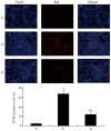
Diabetic nephropathy (DN) is by far the most common cause of end‑stage renal disease (ESRD) in industrial countries, accounting for ~45% of all new ESRD cases in the United States. Grape seed proanthocyanidin extracts (GSPE) are powerful antioxidants, with an antioxidant ability 50‑fold greater than that of vitamin E and 20‑fold greater than that of vitamin C. The present study investigated whether GSPE can protect against streptozotocin (STZ)‑induced DN and aimed to elucidate a possible mechanism. Male Sprague Dawley rats were randomly divided into three groups: Control group (N), diabetes mellitus group (DM) injected with 40 mg/kg STZ, and the GSPE treatment group (intragastric administration of 250 mg/kg/day GSPE for 16 weeks after diabetes was induced in the rats). Blood and kidney samples were collected after treatment. The renal pathological changes were determined with periodic acid‑Schiff (PAS) staining, while the protein expression levels of glucose‑regulated protein 78 (GRP78), phosphorylated‑extracellular signal‑regulated kinase (p‑ERK) and Caspase‑12 were determined by western blotting and immunohistochemical staining. Apoptosis was determined with a terminal deoxynucleotidyl transferase dUTP nick‑end labeling (TUNEL) assay. Compared with the DM group, the GSPE group had no significant changes in the blood urea nitrogen (BUN) level and serum creatinine (Scr) level, but showed a significant decline in the renal index (RI) level and 24‑h urinary albumin level (P<0.05). The histopathology results indicated very little pathological damage in the GSPE group. Compared with the DM group, the GSPE group had a significantly reduced number of TUNEL‑positive cells (P<0.05), and the GSPE group had an obvious reduction in the protein expression of GRP78, p‑ERK, and Caspase‑12 (P<0.05). In this study, the results indicated that GSPE can protect renal function and attenuate endoplasmic reticulum stress‑induced apoptosis via the Caspase‑12 pathway in STZ‑induced DN.
Figures


Similar articles
-
Grape seed proanthocyanidin extract protects from cisplatin-induced nephrotoxicity by inhibiting endoplasmic reticulum stress-induced apoptosis.Mol Med Rep. 2014 Mar;9(3):801-7. doi: 10.3892/mmr.2014.1883. Epub 2014 Jan 3. Mol Med Rep. 2014. PMID: 24399449 Free PMC article.
-
Nephrin loss is reduced by grape seed proanthocyanidins in the experimental diabetic nephropathy rat model.Mol Med Rep. 2017 Dec;16(6):9393-9400. doi: 10.3892/mmr.2017.7837. Epub 2017 Oct 19. Mol Med Rep. 2017. PMID: 29152654 Free PMC article.
-
Thioredoxin is implicated in the anti‑apoptotic effects of grape seed proanthocyanidin extract during hyperglycemia.Mol Med Rep. 2017 Nov;16(5):7731-7737. doi: 10.3892/mmr.2017.7508. Epub 2017 Sep 18. Mol Med Rep. 2017. PMID: 28944891
-
Molecular mechanisms of cardioprotection by a novel grape seed proanthocyanidin extract.Mutat Res. 2003 Feb-Mar;523-524:87-97. doi: 10.1016/s0027-5107(02)00324-x. Mutat Res. 2003. PMID: 12628506 Review.
-
Cellular protection with proanthocyanidins derived from grape seeds.Ann N Y Acad Sci. 2002 May;957:260-70. doi: 10.1111/j.1749-6632.2002.tb02922.x. Ann N Y Acad Sci. 2002. PMID: 12074978 Review.
Cited by
-
Effects of Grape Seed Proanthocyanidin Extract on Obesity.Obes Facts. 2020;13(2):279-291. doi: 10.1159/000502235. Epub 2020 Feb 28. Obes Facts. 2020. PMID: 32114568 Free PMC article.
-
Protective Effects of Grape Seed Proanthocyanidins on the Kidneys of Diabetic Rats through the Nrf2 Signalling Pathway.Evid Based Complement Alternat Med. 2020 Sep 29;2020:5205903. doi: 10.1155/2020/5205903. eCollection 2020. Evid Based Complement Alternat Med. 2020. PMID: 33062013 Free PMC article.
-
Proanthocyanidins alleviate Henoch-Schönlein purpura by mitigating inflammation and oxidative stress through regulation of the TLR4/MyD88/NF-κB pathway.Skin Res Technol. 2024 Sep;30(9):e13921. doi: 10.1111/srt.13921. Skin Res Technol. 2024. PMID: 39252568 Free PMC article.
-
Flavonoids on diabetic nephropathy: advances and therapeutic opportunities.Chin Med. 2021 Aug 7;16(1):74. doi: 10.1186/s13020-021-00485-4. Chin Med. 2021. PMID: 34364389 Free PMC article. Review.
-
Bioactive Compounds, Health Benefits and Food Applications of Grape.Foods. 2022 Sep 7;11(18):2755. doi: 10.3390/foods11182755. Foods. 2022. PMID: 36140883 Free PMC article. Review.
References
MeSH terms
Substances
LinkOut - more resources
Full Text Sources
Other Literature Sources
Medical
Miscellaneous

