. 2018 Aug;18(2):2117-2123.
doi: 10.3892/mmr.2018.9159. Epub 2018 Jun 14.

Amplification of bacterial genomic DNA from all ascitic fluids with a highly sensitive polymerase chain reaction

Affiliations

Amplification of bacterial genomic DNA from all ascitic fluids with a highly sensitive polymerase chain reaction

Hirayuki Enomoto et al. Mol Med Rep. 2018 Aug.

Abstract

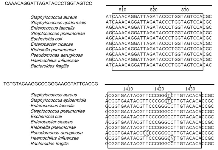
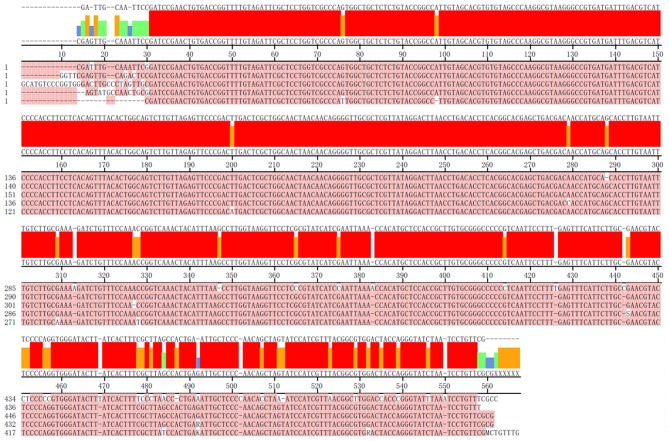
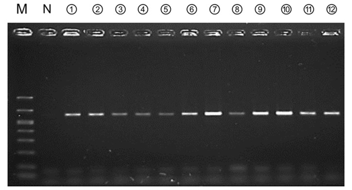
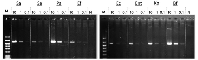
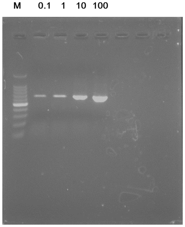
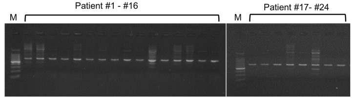
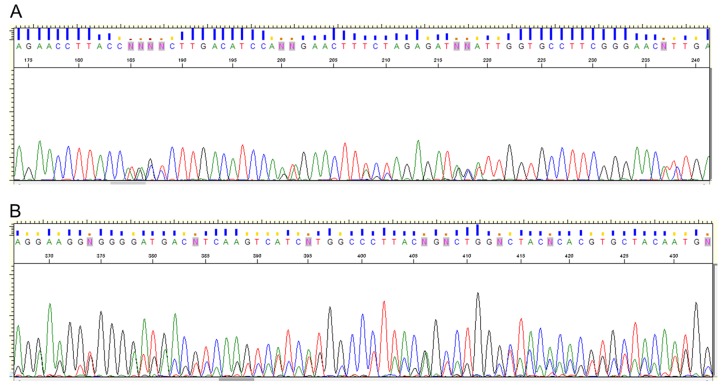
Due to varying positive rates of polymerase chain reaction (PCR) amplification, interpretation of conventional PCR results for non‑infectious ascites remains problematic. The present study developed a highly sensitive PCR protocol and investigated the positive rate of PCR for the 16S ribosomal (r)RNA gene in non‑infectious ascites. Following the design of a new PCR primer pair for the 16S rRNA gene (800F and 1400R), the sequences of PCR products were analyzed and the lower limit for bacterial DNA detection evaluated. The positive rate of PCR for 16S rRNA gene in non‑infectious ascites was also evaluated. PCR with the primer pair amplified the genomic DNA of 16S rRNA genes of major disease‑causing bacterial strains. Additionally, PCR with this primer pair provided highly sensitive detection of bacterial genomic DNA (lower limit, 0.1 pg of template DNA). When DNA samples isolated from ascites were used, the 16S rRNA gene was amplified independently of the presence of bacterial infection. PCR products contained the genomic DNA fragments of multiple bacterial species. Bacterial genomic DNA can be amplified from all ascitic fluids using a highly sensitive PCR protocol. Careful attention is required to interpret the results based on simple amplification of 16S rRNA gene with conventional PCR.


Figures

Figure 1.
Figure 1.
PCR primers corresponding to conserved sequences for multiple bacterial genes. The 800F-primer is a forward primer corresponding to conserved sequences around base pair positions 800. The 1400R-primer is a reverse primer corresponding to conserved sequences around base pair positions 1400 (circle: one base mismatch). PCR, polymerase chain reaction.
Figure 2.
Figure 2.
Sequencing results for PCR products. PCR, polymerase chain reaction.
Figure 3.
Figure 3.
Amplification of the 16S rRNA gene using bacterial genomic DNA samples. PCR products were obtained from DNA samples of various bacterial strains. 1: Escherichia coli; 2: Klebsiella pneumoniae subsp. pneumoniae; 3: Enterobacter cloacae subsp. cloacae; 4: Pseudomonas aeruginosa; 5: Bacteroides fragilis; 6: Staphylococcus aureus; 7: Staphylococcus epidermidis; 8: Enterococcus faecalis; 9: Enterococcus faecium; 10: Streptococcus pneumoniae; 11: Streptococcus pyogenes; 12: Streptococcus agalactiae. PCR, polymerase chain reaction; rRNA, ribosomal RNA; N, Negative control; M, Marker.
Figure 4.
Figure 4.
Amplification of the 16S rRNA gene using a small amount of bacterial DNA templates. With our PCR conditions, evidently detectable bands were obtained with only 0.1 pg of bacterial DNA template. rRNA, ribosomal RNA; Sa, Staphylococcus aureus; Se, Staphylococcus epidermidis; Pa, Pseudomonas aeruginosa; Ef, Enterococcus faecalis; Ec, Escherichia coli; Ent, Enterobacter cloacae subsp. cloacae; Kp, Klebsiella pneumoniae subsp. pneumoniae; Bf, Bacteroides fragilis; N, Negative control; M, Marker.
Figure 5.
Figure 5.
Amplification of 16S rRNA genes with higher efficacy of amplification. When DNA polymerase with higher efficacy of amplification (Prime STAR HS) was used and the annealing temperature of 55°C selected, the 16S rRNA target gene was clearly amplified from the DNA template of Escherichia coli. rRNA, ribosomal RNA.
Figure 6.
Figure 6.
Results of PCRs with DNA samples of non-infectious ascites. PCR bands were obtained for all 24 ascites DNA samples, irrespective of the absence of bacterial infection. M, Marker; PCR, polymerase chain reaction.
Figure 7.
Figure 7.
DNA sequences of PCR with samples of non-infectious ascites. (A) Although PCR products of a patient had homologous to the genomic sequences of 16S rRNA, multi-peak signals were observed at numerous sequence points and identification of specific pathogens was challenging. (B) PCR products with a sample of another patient also showed multi-peak signals at various sequence points.

Similar articles

Cited by

References

    1. Runyon BA. AASLD Practice Guidelines Committee: Management of adult patients with ascites due to cirrhosis: An update. Hepatology. 2009;49:2087–2107. doi: 10.1002/hep.22853. - DOI - PubMed
    1. Garcia-Tsao G, Lim JK. Members of Veterans Affairs Hepatitis C Resource Center Program: Management and treatment of patients with cirrhosis and portal hypertension: recommendations from the Department of Veterans Affairs Hepatitis C Resource Center Program and the National Hepatitis C Program. Am J Gastroenterol. 2009;104:1802–1829. doi: 10.1038/ajg.2009.360. - DOI - PubMed
    1. European Association for the Study of the Liver: EASL clinical practice guidelines on the management of ascites, spontaneous bacterial peritonitis, and hepatorenal syndrome in cirrhosis. J Hepatol. 2010;53:397–417. doi: 10.1016/j.jhep.2010.05.004. - DOI - PubMed
    1. Wiest R, Krag A, Gerbes A. Spontaneous bacterial peritonitis: Recent guidelines and beyond. Gut. 2012;61:297–310. doi: 10.1136/gutjnl-2011-300779. - DOI - PubMed
    1. Enomoto H, Inoue S, Matsuhisa A, Aizawa N, Imanishi H, Saito M, Iwata Y, Tanaka H, Ikeda N, Sakai Y, et al. Development of a new in situ hybridization method for the detection of global bacterial DNA to provide early evidence of a bacterial infection in spontaneous bacterial peritonitis. J Hepatol. 2012;56:85–94. doi: 10.1016/j.jhep.2011.06.025. - DOI - PubMed