. 2018 Aug;18(2):1345-1352.
doi: 10.3892/mmr.2018.9119. Epub 2018 Jun 1.

Telomerase reverse transcriptase induced thyroid carcinoma cell proliferation through PTEN/AKT signaling pathway

Affiliations

Telomerase reverse transcriptase induced thyroid carcinoma cell proliferation through PTEN/AKT signaling pathway

Hao Zhang et al. Mol Med Rep. 2018 Aug.

Abstract

Thyroid carcinoma is the most common endocrine malignant tumor in the world, and so, there is a requirement to develop novel molecular targets for thyroid cancer diagnosis and treatment. Telomerase reverse transcriptase (TERT) was revealed to promote cell proliferation in a number of types of cell. To evaluate whether and how TERT functioned on papillary thyroid cancer (PTC) cell proliferation, the present study constructed TERT over‑expression [recombined (r)TERT plasmid group] and interference [small interfering RNA (si)‑TERT group] models by liposome transfection respectively to study the molecular mechanisms. The transfection efficiency was first detected by reverse transcription‑quantitative polymerase chain reaction (RT‑qPCR) and western blotting to analyze TERT levels compared with the negative control (NC) and control groups. Then MTT and carboxyfluorescein diacetate succinimidyl ester assays were performed to determine living cell proliferation and total cell proliferation respectively. Propidium iodide assay was used to detect alterations in cell cycle progression. RT‑qPCR and western blotting were performed to detect associated factor variation. The results demonstrated that, following the generation of TERT overexpression or silencing PTC cells, the living cells and also total cell proliferation increased significantly in the rTERT group, and decreased significantly in siTERT group, when compared with the NC and control groups. The cell cycle was accelerated in the rTERT group, and blocked in the G1/S transition in the siTERT group. The mRNA and protein levels of P27, P53 and phosphatase and tensin homolog (PTEN) decreased significantly in the rTERP group and increased in the siTERP group, while cyclin dependent kinase 2 and Cyclin D1 increased significantly in the rTERP group and decreased in the siTERP group. The expression of cell division cycle 25A did not alter significantly. The protein levels of β‑catenin and retinoblastoma were also unaltered. Protein kinase B (AKT) was detected once activated by TERT, and there were increased phosphorylated (p)‑AKT protein levels in the rTERT group, and decreased p‑AKT protein levels in the siTERT group. In conclusion, TERT could induce thyroid carcinoma cell proliferation mainly through the PTEN/AKT signaling pathway.


Figures

Figure 1.
Figure 1.
Transfection efficiency of TERT in mixed-PTC cells. (A) Reverse transcription-quantitative polymerase chain reaction was performed to detect the mRNA expression of TERT. TERT expression increased significantly in rTERT group, and decreased significantly in siTERT. (B) Western blotting was performed to detect the protein levels of TERT. (C) TERT protein expression increased significantly in the rTERT group, and decreased significantly in siTERT group, when compared with te NC group. There was no significant difference of TERT expression between NC and control group. Data were presented as mean ± standard deviation (n=3). **P<0.01 vs. NC. TERT, telomerase reverse transcriptase; rTERT, recombined TERT plasmid (overexpression group); siTERT, TERT small interfering RNA (interfering group); NC, negative control.
Figure 2.
Figure 2.
TERT promoted the cell proliferation of mixed-PTC cells. (A) MTT assay was performed to detect the living cell proliferation of mixed-PTC cells. It was indicated that TERT overexpression significantly promoted cell proliferation, and TERT interference markedly inhibited it in time-dependent manner. Cell activity of NC group was not statistically different with the control group. (B) Carboxyfluorescein diacetate succinimidyl ester was used to detect all cell proliferation. (C) The flow cytometry results revealed that, cell proliferation decreased significantly in the siTERT group, and increased significantly in rTERT group. Cell activity of the NC group was not statistically different with the control group. Data were presented as mean ± standard deviation (n=3). **P<0.01 vs. NC. TERT, telomerase reverse transcriptase; rTERT, recombined TERT plasmid (overexpression group); siTERT, TERT small interfering RNA (interfering group); NC, negative control; PTC, papillary thyroid cancer; OD, optical density.
Figure 3.
Figure 3.
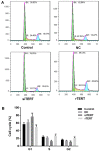
TERT promoted cell cycle progression of mixed-PTC cells. (A) Propidium iodide was used to determine the cell cycle progression of different cells. (B) It demonstrated that TERT interference inhibited the cell proliferation of mixed-PTC cells by blocking the cell cycle from G1/S transition, and TERT overexpression promoted cell proliferation by accelerating cell cycle progression, when compared with the NC group. Cell cycle progression of the NC group was not statistically different with the control group. Data were presented as mean ± standard deviation (n=3). *P<0.05 and **P<0.01 vs. NC. TERT, telomerase reverse transcriptase; rTERT, recombined TERT plasmid (overexpression group); siTERT, TERT small interfering RNA (interfering group); NC, negative control; PTC, papillary thyroid cancer.
Figure 4.
Figure 4.
Expression of TERT regulated cell cycle associated factors in mixed-papillary thyroid cancer cells. (A) Reverse transcription-quantitative polymerase chain reaction and (B) western blotting were conducted to detect the expression of cell cycle associated factors in different groups. (C) Relative protein expression. Data were presented as mean ± standard deviation (n=3). **P<0.01 vs. NC. TERT, telomerase reverse transcriptase; rTERT, recombined TERT plasmid (overexpression group); siTERT, TERT small interfering RNA (interfering group); NC, negative control; PTEN, phosphatase and tensin homolog, protein kinase B; CDK, cyclin dependent kinase; CDC25A, cell division cycle 25A.
Figure 5.
Figure 5.
TERT regulates the AKT signaling pathway in mixed-papillary thyroid cancer cells. (A) Western blotting was performed to evaluate the (B) protein levels of Rb, β-catenin p-AKT and AKT. Data were presented as mean ± standard deviation (n=3). **P<0.01 vs. NC. TERT, telomerase reverse transcriptase; rTERT, recombined TERT plasmid (overexpression group); siTERT, TERT small interfering RNA (interfering group); NC, negative control; p-, phosphorylated; AKT, protein kinase B; Rb, retinoblastoma.

Similar articles

Cited by

References

    1. Davies L, Welch HG. Increasing incidence of thyroid cancer in the United States, 1973–2002. JAMA. 2006;295:2164–2167. doi: 10.1001/jama.295.18.2164. - DOI - PubMed
    1. Wang Y, Wang W. Increasing incidence of thyroid cancer in Shanghai, China, 1983–2007. Asia Pac J Public Health. 2015;27:NP223–NP229. doi: 10.1177/1010539512436874. - DOI - PubMed
    1. Albores-Saavedra J, Henson DE, Glazer E, Schwartz AM. Changing patterns in the incidence and survival of thyroid cancer with follicular phenotype-papillary, follicular, and anaplastic: A morphological and epidemiological study. Endocr Pathol. 2007;18:1–7. doi: 10.1007/s12022-007-0002-z. - DOI - PubMed
    1. Wynford-Thomas D. Origin and progression of thyroid epithelial tumours: Cellular and molecular mechanisms. Horm Res. 1997;47:145–157. doi: 10.1159/000185458. - DOI - PubMed
    1. Gomez DE, Armando RG, Farina HG, Menna PL, Cerrudo CS, Ghiringhelli PD, Alonso DF. Telomere structure and telomerase in health and disease (review) Int J Oncol. 2012;41:1561–1569. doi: 10.3892/ijo.2012.1611. - DOI - PMC - PubMed