Review
. 2018 Jul;44(4):231-247.
doi: 10.1097/ICL.0000000000000499.

A Review of Current Concepts of the Etiology and Treatment of Myopia

Affiliations
Review

A Review of Current Concepts of the Etiology and Treatment of Myopia

Jeffrey Cooper et al. Eye Contact Lens. 2018 Jul.

Abstract

Myopia occurs in more than 50% of the population in many industrialized countries and is expected to increase; complications associated with axial elongation from myopia are the sixth leading cause of blindness. Thus, understanding its etiology, epidemiology, and the results of various treatment regiments may modify current care and result in a reduction in morbidity from progressive myopia. This rapid increase cannot be explained by genetics alone. Current animal and human research demonstrates that myopia development is a result of the interplay between genetic and the environmental factors. The prevalence of myopia is higher in individuals whose both parents are myopic, suggesting that genetic factors are clearly involved in myopia development. At the same time, population studies suggest that development of myopia is associated with education and the amount time spent doing near work; hence, activities increase the exposure to optical blur. Recently, there has been an increase in efforts to slow the progression of myopia because of its relationship to the development of serious pathological conditions such as macular degeneration, retinal detachments, glaucoma, and cataracts. We reviewed meta-analysis and other of current treatments that include: atropine, progressive addition spectacle lenses, orthokeratology, and multifocal contact lenses.


Conflict of interest statement

J. Cooper: Consultant to VTI (Visioneering Technologies, Inc., Alpharetta, GA), Treehouse Eyes, and Magic Leap, the remaining author has no funding or conflicts of interest to disclose.

Figures

FIG. 1.
FIG. 1.
Regional blur causes axial elongation. Regional retinal blur created in half the retina causes regional elongation of the eye. This occurs even when the optic nerve is cut, but will not occur if atropine is injected into the eye. The eye recognizes the direction of the blur, that is, plus or minus lenses and the region of retinal blur. Reprinted with permission from Cooper J, Schulman E, Jamal N. Current status on the development and treatment of myopia. Optometry 2012;83:179–99.
FIG. 2.
FIG. 2.
Image shells on the retina. Once the eye elongates in myopia, optical images from spherical lenses no longer fall on the retinal plane. The peripheral images are out of focus falling on a plane behind the retina. It is thought that the relative hyperopic error created is the stimulus for axial elongation. Current optical treatments move the peripheral focus in front of the retina.
FIG. 3.
FIG. 3.
Risk of ocular disease with increased myopia. It is readily apparent that the risk of retinal detachment and macular degeneration increases logarithmically with the increase of acquired myopia.139 The risk begins with as little as 1.00 D of myopia. Reprinted with permission from Flitcroft DI. The complex interactions of retinal, optical and environmental factors in myopia aetiology. Prog Retin Eye Res 2012;31:622–60.
FIG. 4.
FIG. 4.
Meta-analysis of progressives and bifocal spectacle lenses. Meta-analysis of 9 clinical trials in which progressive additional and bifocal spectacle lenses (MFL) are compared with single-vision lenses (SVL) using spherical equivalent (A) and axial length (B). Mean difference between SVL and MFL was 0.25 D per year and in those that reported axial length changes, the difference was 0.012 mm.,– The benefit of MFL was greater in Asian versus white children (0.32 D vs. 0.10 D) and/or those that initially had a higher baseline refraction. (Less than 3 D at baseline = 0.16 D vs. greater than 3 D at baseline 0.39 D). It should be noted that these findings were not replicated in an analysis of 16 treatment protocols for myopia. Reprinted with permission from Li SM, Ji YZ, Wu SS, et al. Multifocal versus school-age children: a meta-analysis. Surv Ophthalmol 2011;56:451–60.
FIG. 5.
FIG. 5.
Meta-analysis of orthokeratology. Meta-analysis of 7 OK studies, was performed, which included 435 subjects who were aged between 6 and 16 years.,,,,– Meta analysis found a mean difference between controls and OK patients of 0.26 mm over 2 years. This is a 40% reduction in the progression of myopia. Reprinted with permission from Si JK, Tang K, Bi HS, Guo DD, Guo JG, Wang XR. Orthokeratology for myopia control: A meta-analysis. Optom Vis Sci 2015.
FIG. 6.
FIG. 6.
FIG. 6. Meta-analysis of multifocal contact lenses. Meta-analysis, which included eight studies published between 1999 and 2016, that compared single-vision soft lenses with both concentric ring bifocal soft contact lenses (CCML),,– and peripheral add soft contact lenses (MCL).,, There was less myopia progression with both lenses (the CCML had a weighted mean difference [WMD] of 0.31 D and reduced axial elongation WMD of –0.12 mm, whereas MCL had a WMD of 0.22 D and less axial elongation of 0.10 at the end of 1 year). This represented a 31% reduction of progression with the CCML and 51% reduction with MCL. Axial length reduction was also noted: 38% with the CCML and 51% with MCL after 2 years. Reprinted with permission from Li SM, Kang MT, Wu SS, et al. Studies using concentric ring bifocal and peripheral add multifocal contact lenses to slow myopia progression in school-aged children: a meta-analysis. Ophthalmic Physiol Opt 2016.
FIG. 7.
FIG. 7.
Atropine 1% versus control in slowing myopic progression. Data from the ATOM 1 study are pictorially presented and clearly show the effectivity of atropine over control. Seventy percent of the atropine subjects had less than 0.5 D of progression compared with less than 20% of the controls. It is apparent that atropine 1% results in strong control of myopia progression. Reprinted with permission from Cooper J, Schulman E, Jamal N. Current status on the development and treatment of myopia. Optometry 2012;83:179–99.
FIG. 8.
FIG. 8.
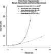
Myopic progression with various doses of atropine. Shih et al. demonstrated that the ability of atropine to control progression is directly related to concentration. The higher the dosage, the more effective atropine is in slowing the progression of myopia. It is clear that even at a relatively low dosage of atropine 0.01%, there is a clinically effective retardation of the progression of myopia. Reprinted with permission from Shih YF, Chen CH, Chou AC, Ho TC, Lin LL, Hung PT. Effects of different concentrations of atropine on controlling myopia in myopic children. J Ocul Pharmacol Ther 1999;15:85–90.
FIG. 9.
FIG. 9.
Progression of myopia during 3 phases of ATOM studies. This graph depicts the cycloplegic refractions (spherical equivalent) in all 3 phases of the ATOM 1 and 2 studies. The first phase was for 2 years during which subjects were randomized to receive various concentrations of atropine (1%, 0.5%). After 2 years, treatment was stopped in all groups for 1 year of time. Those patients still showing more than 0.50 diopters of myopia progression were placed on atropine 0.01% and followed for another 2 years. Reprinted with permission from Chia A, Lu QS, Tan D. Five-year clinical trial on atropine for the treatment of myopia 2: Myopia control with atropine 0.01% eyedrops. Ophthalmology 2016;123:391–99.
FIG. 10.
FIG. 10.
Changes in AL and SPH EQ after 2 years of treatment. Figure 10 depicts the changes in axial length in millimeters (yellow bars going up); spherical equivalent in diopters calculated from the axial length data (red going down); and cycloplegic automated refractor measurements in diopters (green going down) at the end of the 24-month treatment period. The measurements were derived from the ATOM 1 study for atropine 1% and placebo and ATOM 2 for atropine 0.01%, 0.1%, and 0.5%, respectively. It is readily apparent that there is no real difference between axial length measurements after 24 months between placebo and atropine 0.01%; moderate changes with atropine 0.1% and 0.5%; and dramatic changes with atropine 1% (yellow bars). However, the spherical equivalent measurements (green bars), compared with placebo in diopters, show a much greater change over time again being greatest for atropine 1%. The difference between the effect of atropine 0.01% and atropine 1% is not nearly as great as the concentration differences.
FIG. 11.
FIG. 11.
Meta-analysis of 16 different treatments. A meta-analysis of 16 different treatments for myopia was performed using a comparison with either placebo or single-vision spectacle lenses with the following: high-dose atropine (refraction change: 0.68; axial length change –0.21); moderate-dose atropine (refraction change: 0.53; axial length change: –0.21); low-dose atropine (refraction change: 0.53 axial length change: –0.15); pirenzepine (refraction change: 0.29; axial length change: –0.09); OK (axial length change: –0.15); multifocal contact lenses (axial length change: –0.11); and progressive-addition spectacle lenses (refraction change: 0.14 axial length change). Reprinted with permission from Huang J, Wen D, Wang Q, et al. Efficacy comparison of 16 interventions for myopia control in children: A network meta-analysis. Ophthalmology 2016;123:697–708.
FIG. 12.
FIG. 12.
Percentage of reduction of myopia progression with various treatments. Calculated percentage of reduction of progression of myopia for each treatment. Meta-analysis numbers were used to calculate the reduction in progression. Meta-analysis only included prospective clinical trials. Cooper et al. previously calculated the reduction in progression from all published studies without regard to methodology.

References

    1. Flitcroft DI. The complex interactions of retinal, optical and environmental factors in myopia aetiology. Prog Retin Eye Res 2012;31:622–660. - PubMed
    1. Holden BA. The Charles F. Prentice award Lecture 2014: A 50-year research journey: Giants and Great Collaborators. Optom Vis Sci 2015;92:741–749. - PubMed
    1. Tkatchenko AV, Tkatchenko TV, Guggenheim JA, et al. APLP2 regulates refractive error and myopia development in mice and humans. PLoS Genet 2015;11:e1005432. - PMC - PubMed
    1. Pararajasegaram R. VISION 2020-the right to sight: From strategies to action. Am J Ophthalmol 1999;128:359–360. - PubMed
    1. Kempen JH, Mitchell P, Lee KE, et al. The prevalence of refractive errors among adults in the United States, Western Europe, and Australia. Arch Ophthalmol 2004;122:495–505. - PubMed