

Ovarian Carcinoma Histotype: Strengths and Limitations of Integrating Morphology With Immunohistochemical Predictions
- PMID: 29901523
- PMCID: PMC6291360
- DOI: 10.1097/PGP.0000000000000530
Ovarian Carcinoma Histotype: Strengths and Limitations of Integrating Morphology With Immunohistochemical Predictions
Abstract
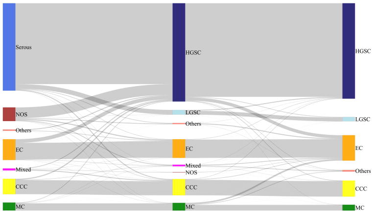
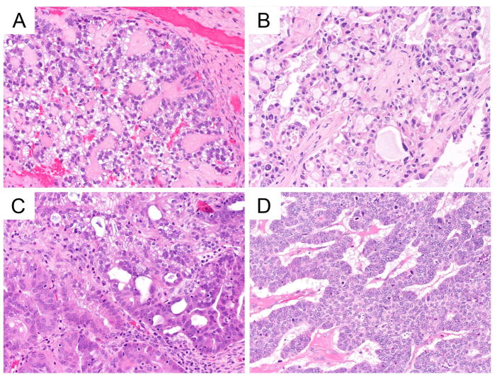
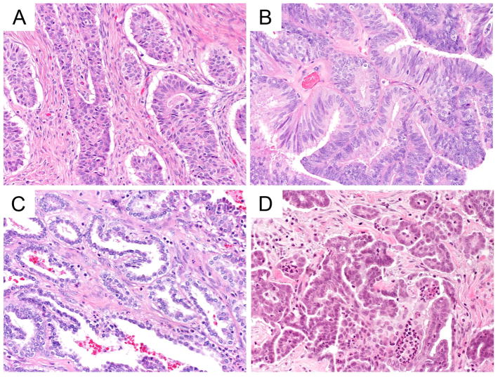
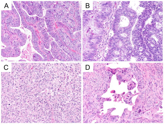
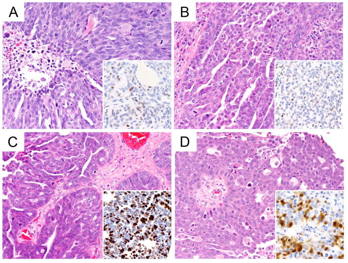
Ovarian carcinoma histotypes are critical for research and patient management and currently assigned by a combination of histomorphology +/- ancillary immunohistochemistry (IHC). We aimed to validate the previously described IHC algorithm (Calculator of Ovarian carcinoma Subtype/histotype Probability version 3, COSPv3) in an independent population-based cohort, and to identify problem areas for IHC predictions. Histotype was abstracted from cancer registries for eligible ovarian carcinoma cases diagnosed from 2002 to 2011 in Alberta and British Columbia, Canada. Slides were reviewed according to World Health Organization 2014 criteria, tissue microarrays were stained with and scored for the 8 COSPv3 IHC markers, and COSPv3 histotype predictions were calculated. Discordant cases for review and COSPv3 prediction were arbitrated by integrating morphology with IHC results. The integrated histotype (N=880) was then used to identify areas of inferior COSPv3 performance. Review histotype and integrated histotype demonstrated 93% agreement suggesting that IHC information revises expert review in up to 7% of cases. There was also 93% agreement between COSPv3 prediction and integrated histotype. COSPv3 errors predominated in 4 areas: endometrioid carcinoma (EC) versus clear cell (N=23), EC versus low-grade serous (N=15), EC versus high-grade serous (N=11), and high-grade versus low-grade serous (N=6). Most problems were related to Napsin A-negative clear cell, WT1-positive EC, and p53 IHC wild-type high-grade serous carcinomas. Although 93% of COSPv3 prediction accuracy was validated, some histotyping required integration of morphology with ancillary test results. Awareness of these limitations will avoid overreliance on IHC and misclassification of histotypes for research and clinical management.
Conflict of interest statement
The authors declare no conflict of interest.
Figures
References
-
- Mirza MR, Monk BJ, Herrstedt J, Oza AM, Mahner S, Redondo A, et al. Niraparib Maintenance Therapy in Platinum-Sensitive, Recurrent Ovarian Cancer. The New England journal of medicine. 2016;375(22):2154–64. - PubMed
-
- Lemery S, Keegan P, Pazdur R. First FDA Approval Agnostic of Cancer Site - When a Biomarker Defines the Indication. The New England journal of medicine. 2017;377(15):1409–12. - PubMed
-
- Rambau PF, Duggan MA, Ghatage P, Warfa K, Steed H, Perrier R, et al. Significant frequency of MSH2/MSH6 abnormality in ovarian endometrioid carcinoma supports histotype-specific Lynch syndrome screening in ovarian carcinomas. Histopathology. 2016;69(2):288–97. - PubMed
MeSH terms
Substances
Grants and funding
LinkOut - more resources
Full Text Sources
Other Literature Sources
Medical
Research Materials
Miscellaneous
