Clinical Trial
. 2018 Jun;97(24):e11095.
doi: 10.1097/MD.0000000000011095.

Curcumin induces endoplasmic reticulum stress-associated apoptosis in human papillary thyroid carcinoma BCPAP cells via disruption of intracellular calcium homeostasis

Affiliations
Clinical Trial

Curcumin induces endoplasmic reticulum stress-associated apoptosis in human papillary thyroid carcinoma BCPAP cells via disruption of intracellular calcium homeostasis

Li Zhang et al. Medicine (Baltimore). 2018 Jun.

Abstract

Background: Thyroid cancer is the most common endocrine tumor. Our previous studies have demonstrated that curcumin can induce apoptosis in human papillary thyroid carcinoma BCPAP cells. However, the underlined mechanism has not been clearly elucidated. Endoplasmic reticulum (ER) is a major organelle for synthesis, maturation, and folding proteins as well as a large store for Ca. Overcoming chronically activated ER stress by triggering pro-apoptotic pathways of the unfolded protein response (UPR) is a novel strategy for cancer therapeutics. Our study aimed to uncover the ER stress pathway involved in the apoptosis caused by curcumin.

Methods: BCPAP cells were treated with different doses of curcumin (12.5-50 μM). Annexin V/PI double staining was used to determine cell apoptosis. Rhod-2/AM calcium fluorescence probe assay was performed to measure the calcium level of endoplasmic reticulum. Western blot was used to examine the expression of ER stress marker C/EBP homologous protein 10 (CHOP) and glucose-regulated protein 78 (GRP78). X-box binding protein1 (XBP-1) spliced form was examined by reverse transcriptase-polymerase chain reaction (RT-PCR).

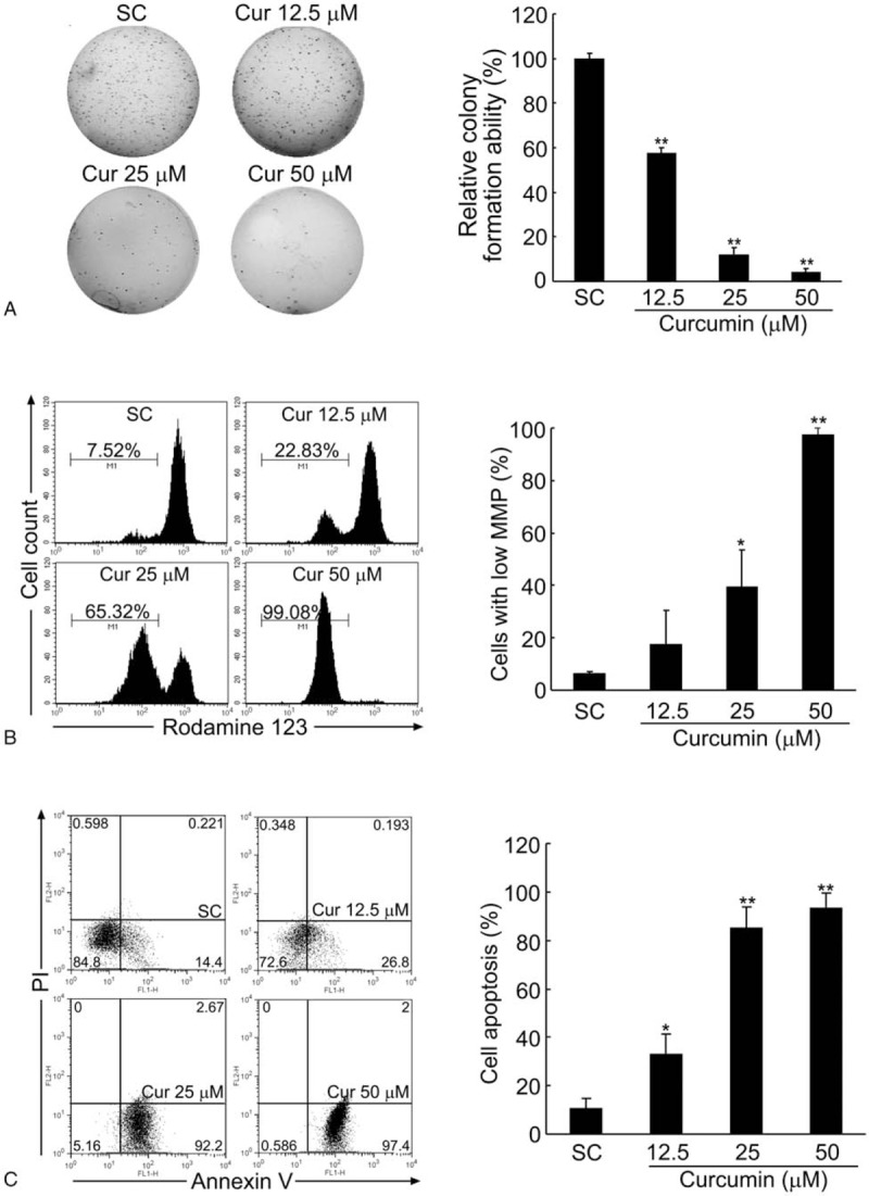
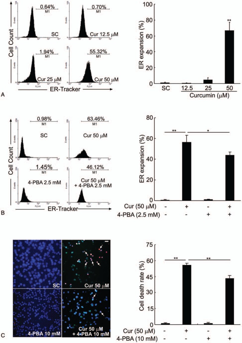
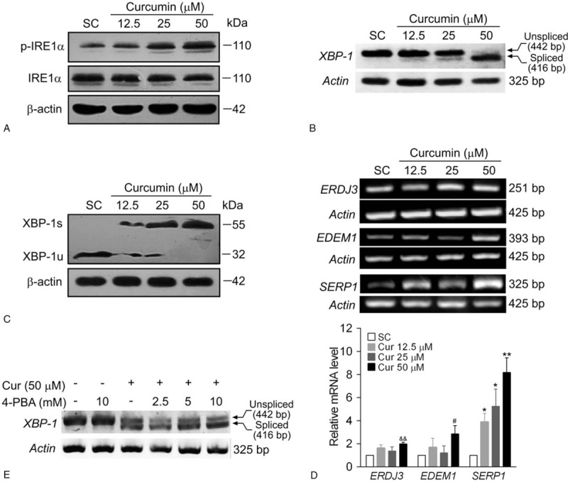
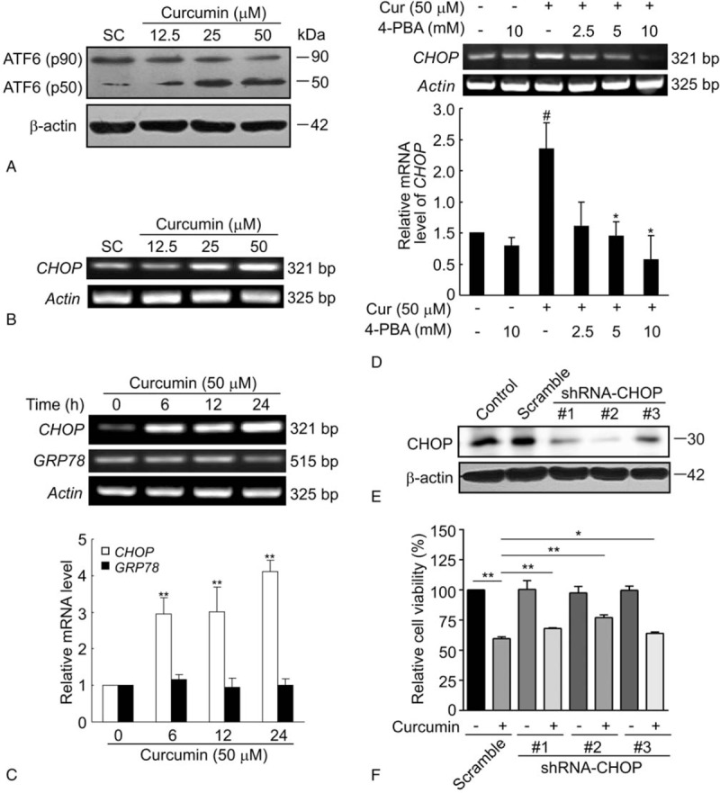
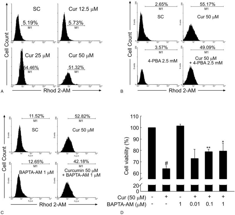
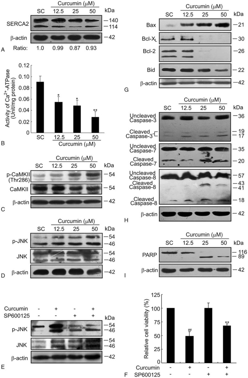
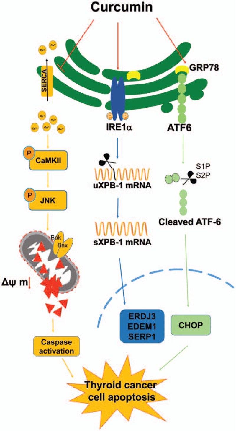
Results: Curcumin significantly inhibited anchorage-independent cell growth and induced apoptosis in BCPAP cells. Curcumin induced ER stress and UPR responses in a dose- and time-dependent manner, and the chemical chaperone 4-phenylbutyrate (4-PBA) partially reversed the antigrowth activity of curcumin. Moreover, curcumin significantly increased inositol-requiring enzyme 1α (IRE1α) phosphorylation and XBP-1 mRNA splicing to induce a subsets of ER chaperones. Increased cleavage of activating transcription factor 6 (ATF6), which enhances expression of its downstream target CHOP was also observed. Furthermore, curcumin induced intracellular Ca influx through inhibition of the sarco-endoplasmic reticulum ATPase 2A (SERCA2) pump. The increased cytosolic Ca then bound to calmodulin to activate calcium/calmodulin-dependent protein kinase II (CaMKII) signaling, leading to mitochondrial apoptosis pathway activation. Ca chelator BAPTA partially reversed curcumin-induced ER stress and growth suppression, confirming the possible involvement of calcium homeostasis disruption in this response.

Conclusions: Curcumin inhibits thyroid cancer cell growth, at least partially, through ER stress-associated apoptosis. Our observations provoked that ER stress activation may be a promising therapeutic target for thyroid cancer treatment.(Figure is included in full-text article.).


Conflict of interest statement

The authors declare that there are no conflicts of interest.

Figures

Figure 1
Figure 1
Curcumin inhibits the colony formation and induces the apoptosis of BCPAP cells. (A) Curcumin inhibits the colony formation ability of BCPAP cells in a dose-dependent manner. BCPAP cells were treated with different doses (12.5–50 μM) of curcumin, and the colony formation ability was measured by soft agar assay. (B) Curcumin decreases the mitochondrial membrane potential (MMP) of BCPAP cells. After treated by different doses (12.5–50 μM) of curcumin for 24 hours, MMP of BCPAP cells were determined by flow cytometry using Rhodamine 123 fluorescent dye. (C) Curcumin induces the apoptosis of BCPAP cells. BCPAP cells were treated with different doses (12.5–50 μM) of curcumin for 24 hours, and the apoptotic cells were determined by Annexin V-FITC/PI analysis. Percentage of living cells (lower left: Annexin V-FITC/PI), early apoptosis cells (lower right: Annexin V-FITC+/PI), and late apoptosis cells (upper right: Annexin V-FITC+/PI+) were showed in the flow cytometry chart. All data shown represent the means ± S.D. of 3 independent experiments. SC =  solvent control. ∗P < .05, ∗∗P < .01.
Figure 2
Figure 2
Curcumin induces ER expansion in BCPAP cells. (A) Curcumin enlarges ER size in BCPAP cells. After treatment with different doses of curcumin (12.5–50 μM), the ER size of BCPAP cells was measured by flow cytometry analysis using the ER-Tracker dyes. (B) ER stress inhibitor, 4-PBA, partially reverses the ER expansion induced by curcumin in BCPAP cells. Cells were pretreated with or without 2.5 mM of 4-PBA for 1 hour, followed by exposure to 50 μM of curcumin for another 24 hours. The degree of ER expansion was detected by flow cytometry. (C) 4-PBA ameliorates the cell death induced by curcumin. BCPAP cells were pretreated with or without 10 mM of 4-PBA for 1 hour before incubation with 50 μM of curcumin for another 24 hours. Representative images of Hoechst/PI staining were shown and white arrows indicated PI-positive cells. Scale bar, 10 μm. The cell death rate is expressed as percentage of PI-positive staining cells. All data shown represent the means ± S.D. of 3 independent experiments. SC = solvent control. ∗P < .05, ∗∗P < .01.
Figure 3
Figure 3
Curcumin induces phosphorylation of IRE1α and XBP-1 mRNA splicing. BCPAP cells were exposed to different dosages (12.5–50 μM) of curcumin for 24 hours. After the cells were collected, western blot or RT-PCR analysis were performed. (A) Curcumin increases the phosphorylation of IRE1α in BCPAP cells. The protein levels of phosphorylated IRE1α and total IRE1α were detected by western blot analysis. β-actin was used as a loading control. (B) Curcumin increases XBP-1 splicing in BCPAP cells. The mRNA levels of spliced and unspliced forms of XBP-1 were assessed by RT-PCR. Actin was performed as a loading control. (C) Curcumin treatment results in the conversion of inactive unspliced XBP-1 (XBP-1u) protein to an active spliced (XBP-1s) protein in BCPAP cells. The protein levels of spliced and unspliced forms of XBP-1 were detected by western blot assay. (D) Curcumin enhances the mRNA expressions of XBP-1 downstream genes. BCPAP cells were exposed to different dosages (12.5–50 μM) of curcumin for 24 hours. The mRNA expressions of ERDJ3, EDEM1, and SERP1 were determined by RT-PCR and normalized to that of Actin. The data shown represent the means ± S.D. of 3 independent experiments. &&P < .01, #P < .05, ∗P < .05, ∗∗P < .01 versus SC group. (E) 4-PBA fails to rescue the XBP-1 splicing induced by curcumin in BCPAP cells. Cells were pretreated with or without different dosages of 4-PBA (2.5–10 mM) for 1 hour. After that, cells were treated with 50 μM of curcumin for 24 hours and XBP-1 splicing was measured by RT-PCR. IRE1α = inositol-requiring enzyme 1α, RT-PCR = reverse transcriptase-polymerase chain reaction.
Figure 4
Figure 4
Curcumin elevates ATF6 cleavage and CHOP mRNA expression. (A) Curcumin induces the cleavage of ATF6 in BCPAP cells. Cells were treated with different dosages (12.5–50 μM) of curcumin for 24 hours and the uncleaved (90 kDa) and cleaved (50 kDa) forms of ATF6 were detected by western blot assay. (B) Curcumin dose-dependently increases the CHOP mRNA expression of BCPAP cells. BCPAP cells were exposed to different dosages (12.5–50 μM) of curcumin for 24 hours and subjected to RT-PCR analysis. (C) Curcumin increases the CHOP mRNA expression of BCPAP cells in a time-dependent manner. BCPAP cells were treated with 50 μM of curcumin for indicated times. Total RNAs were isolated and subjected to RT-PCR. CHOP and GRP78 mRNA expression were determined and normalized to that of Actin. (D) 4-PBA reverses the upregulation of CHOP mRNA induced by curcumin. BCPAP cells were pretreated with or without different dosages (2.5–10 μM) of 4-PBA before 50 μM of curcumin exposure for additional 24 hours. CHOP mRNA expression was detected by RT-PCR. All data shown represent the means ± S.D. of 3 independent experiments. #P < .05 versus untreated group; ∗P < .05, ∗∗P < .01 versus curcumin alone-treated group. (E) BCPAP cells were transfected with 3 siRNAs targeting different region of CHOP mRNA (shRNA-CHOP #1, #2, and #3) or negative control (Scramble). Cell lysates were collected 24 hours later and CHOP knockdown efficiency were determined by western blot. (F) BCPAP cells were transfected with shRNA-CHOP #1, #2, and #3 or scramble control. After 24 hours, cells were treated with or without 50 μM of curcumin for another 24 hours. The cell survival rate was determined by MTT assay. ∗P < .05, ∗∗P < .01. ATF6 = activating transcription factor 6.
Figure 5
Figure 5
Curcumin increases the intracellular Ca2+ level of BCPAP cells. (A) Curcumin increases the intracellular Ca2+ content in BCPAP cells. After BCPAP cells were exposed to different dosages (12.5–50 μM) of curcumin for 24 hours, intracellular Ca2+ level was detected by flow cytometry analysis using Rhod 2-AM. (B) 4-PBA fails to rescue the increased intracellular Ca2+ content induced by curcumin. After treatment with 50 μM curcumin, 2.5 mM 4-PBA, or the combination of 50 μM curcumin and 2.5 mM 4-PBA, the intracellular Ca2+ content of BCPAP cells was determined by Flow cytometry. (C) BAPTA-AM decreases the elevated intracellular Ca2+ level induced by curcumin. BCPAP cells were preincubated in the presence or absence of BAPTA-AM (1 μM) for 0.5 hour followed by curcumin (50 μM) treatment for 24 hours. After incubation, the intracellular Ca2+ content of BCPAP cells was determined by flow cytometry. (D) BAPTA-AM protects BCPAP cells from curcumin-induced cytotoxicity. Cells were treated as in (C) and the cell viability of BCPAP cells was measured by MTT assay. All data shown represent the means ± S.D. of 3 independent experiments. #P < .05 versus untreated group; ∗P < .05, ∗∗P < .01 versus curcumin alone-treated group.
Figure 6
Figure 6
Curcumin promotes apoptosis by activating Ca2+-CaMKII-JNK pathway. (A) Curcumin slightly decreases the protein level of SERCA2 in BCPAP cells. BCPAP cells were exposed to different dosages (12.5–50 μM) of curcumin for 24 hours and the protein level of SERCA2 was detected by western blotting. β-actin was used as a loading control. Relative protein levels of SERCA2 were shown at the bottom of the graph. (B) Curcumin inhibits SERCA2 activity of BCPAP cells. Cells were incubated with various concentrations of curcumin for 24 hours. Then the cells were harvested and the Ca2+-ATPase activity was measured according to the instructions of the Ca2+-ATPase detection kit. Error bars are means ± S.D. of 3 independent experiments. ∗P < .05, ∗∗P < .01 versus SC group. (C) Curcumin treatment increases the phosphorylation of CaMKII in BCPAP cells. (D) Curcumin increases the phosphorylation of JNK of BCPAP cells. (E) BCPAP cells were either treated with 50 μM of curcumin for 24 hours or pre-treated with 10 μM of SP600125 for 1 hour before curcumin treatment. The phosphorylated and total JNK protein levels were detected by western blot assay. β-actin was used as a loading control. (F) BCPAP cells were treated as in (E). MTT assay was used to determine the cell viability. ##P < .01 versus untreated group; ∗∗P < .01 versus curcumin alone-treated group. (G) Curcumin induces apoptosis of BCPAP cells. BCPAP cells were treated with curcumin at different dosages for 24 hours and then harvested for apoptosis analysis. The pro-apoptotic protein Bax and anti-apoptotic protein Bcl-XL, Bcl2, Bid were measured by western blotting. (H) Curcumin activates caspase-3, -7, and -8 pathway in BCPAP cells. (I) Curcumin treatment leads to an increased cleavage of PARP. These apoptosis-related proteins were determined at least 3 times and representative data were shown. PARP = poly ADP-ribose polymerase; SC = solvent control.
None

Similar articles

Cited by

References

    1. Grande E, Diez JJ, Zafon C, et al. Thyroid cancer: molecular aspects and new therapeutic strategies. J Thyroid Res 2012;2012:847108. - PMC - PubMed
    1. Carneiro RM, Carneiro BA, Agulnik M, et al. Targeted therapies in advanced differentiated thyroid cancer. Cancer Treat Rev 2015;41:690–8. - PubMed
    1. Heger M, van Golen RF, Broekgaarden M, et al. The molecular basis for the pharmacokinetics and pharmacodynamics of curcumin and its metabolites in relation to cancer. Pharmacol Rev 2014;66:222–307. - PubMed
    1. Wang L, Zhang B, Huang F, et al. Curcumin inhibits lipolysis via suppression of ER stress in adipose tissue and prevents hepatic insulin resistance. J Lipid Res 2016;57:1243–55. - PMC - PubMed
    1. Basile V, Belluti S, Ferrari E, et al. bis-Dehydroxy-Curcumin triggers mitochondrial-associated cell death in human colon cancer cells through ER-stress induced autophagy. PLoS One 2013;8:e53664. - PMC - PubMed

Publication types

MeSH terms