

Surgery combined with radio-chemotherapy for esophageal mucoepidermoid carcinoma: A case report
- PMID: 29901650
- PMCID: PMC6023669
- DOI: 10.1097/MD.0000000000011165
Surgery combined with radio-chemotherapy for esophageal mucoepidermoid carcinoma: A case report
Abstract
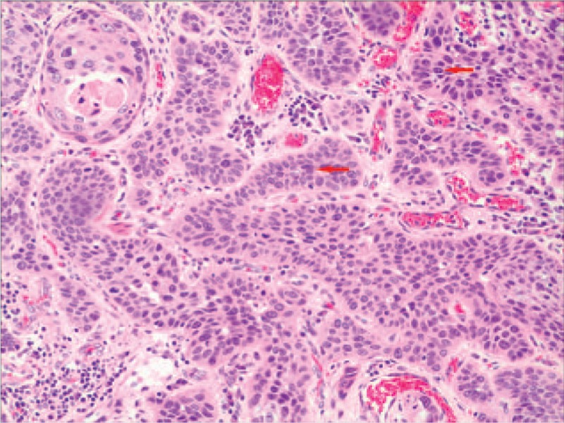
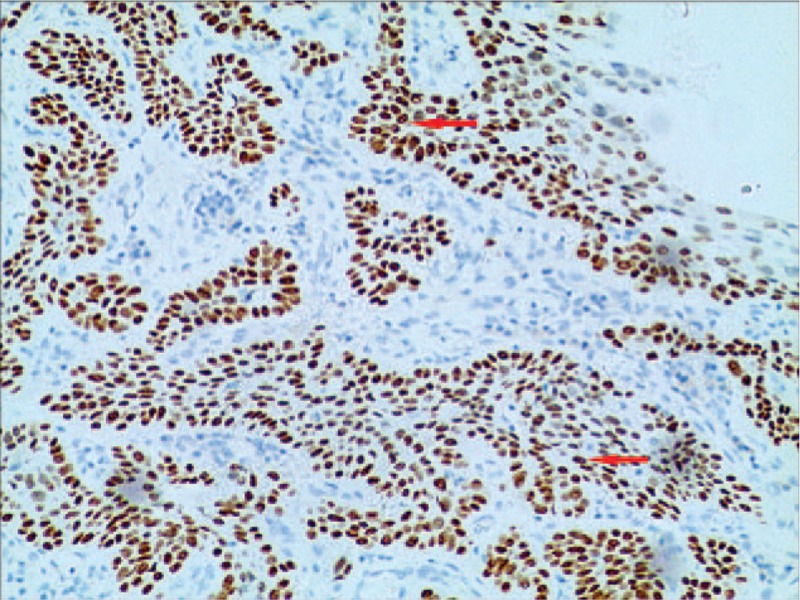
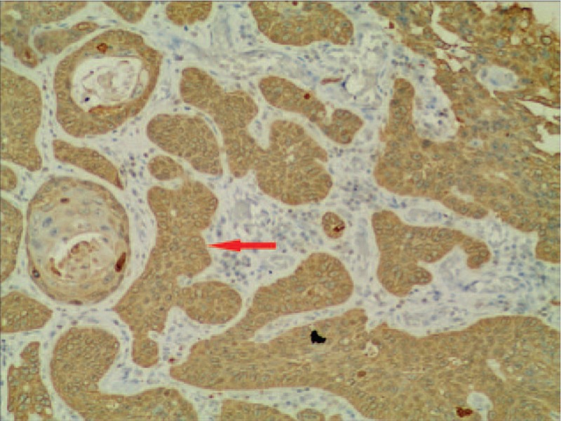
Rationale: Primary mucoepidermoid carcinoma (MEC) of the esophagus is a rare type of malignant neoplasm. Its morphology resembles that of MEC of the salivary glands. It is characterized by a diffuse mixture of squamous and mucus-secreting glandular carcinoma cells. Due to the low incidence of esophageal MEC, the biological behavior and treatment of this tumor have not been well studied.
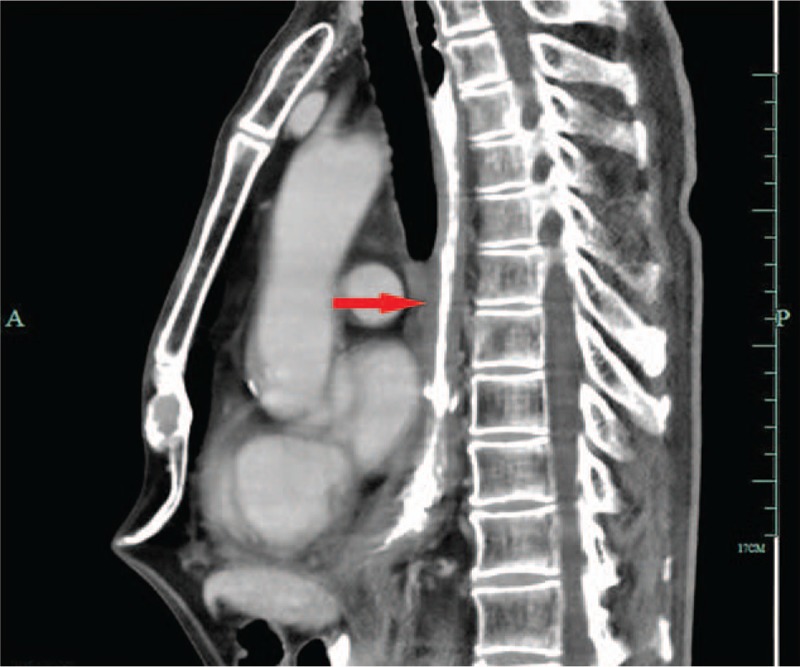
Patient concerns: In this case report, we describe a case of a 59-year-old man who presented with difficulty in swallowing. Iohexol swallowing revealed a malignant-appearing structure in the inferior-thoracic region.
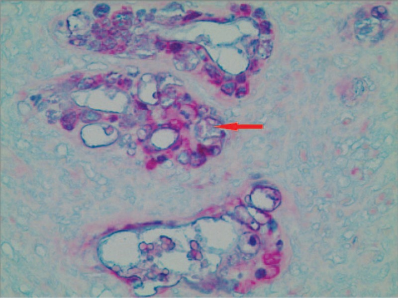
Diagnoses: Biopsy of the lesion under endoscopy demonstrated a mucoepidermoid carcinoma of the esophagus.
Interventions: We performed esophagectomy, esophagogastrostomy through the esophageal bed and 2-field lymphadenectomy. Histopathological analysis of the tumor revealed histological characteristics typical of an esophageal MEC. Radio-chemotherapy was administered to this patient.
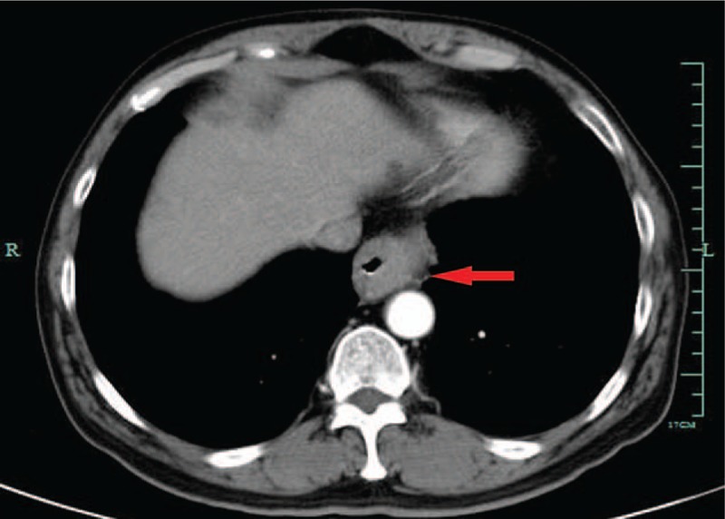
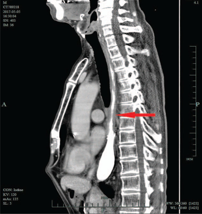
Outcomes: Seventeen months after surgery, an esophageal computed tomography (CT) scan revealed that the wall of esophagus was evenly thickened. However, endoscopic assessment revealed no evidence of recurrence. Further CT scans at 19 and 31 months after surgery also showed a thickened esophageal wall, although endoscopic assessment at 31 months still revealed no esophageal stricture and no evidence of recurrence. The patient is alive with no dysphagia and no evidence of recurrence for over 39 months.
Lessons: There is little evidence of effective treatment nor guidelines for treatment of esophageal MEC. Although the general prognosis of esophageal MEC is poor, comprehensive treatment of surgery and radio-chemotherapy appeared to be effective in this case. Radio-chemotherapy is a possible treatment option that was shown to have acceptable short-term effects.
Conflict of interest statement
The authors declare that they have no competing interests.
Figures
Similar articles
-
A rare case of primary mucoepidermoid carcinoma of the esophagus.Clin J Gastroenterol. 2015 Feb;8(1):26-8. doi: 10.1007/s12328-014-0546-7. Epub 2014 Dec 5. Clin J Gastroenterol. 2015. PMID: 25475139
-
Primary mucoepidermoid carcinoma of the esophagus.J Thorac Oncol. 2011 Aug;6(8):1426-31. doi: 10.1097/JTO.0b013e31821cfb96. J Thorac Oncol. 2011. PMID: 21587086
-
Mucoepidermoid carcinoma of esophagus combined with squamous carcinoma of lung: A case report and literature review.J Cancer Res Ther. 2015 Jul-Sep;11(3):658. doi: 10.4103/0973-1482.140986. J Cancer Res Ther. 2015. PMID: 26458663 Review.
-
[Clinical analysis of 36 cases of esophageal mucoepidermoid carcinoma].Zhonghua Zhong Liu Za Zhi. 2012 Mar;34(3):232-5. doi: 10.3760/cma.j.issn.0253-3766.2012.03.016. Zhonghua Zhong Liu Za Zhi. 2012. PMID: 22780981 Chinese.
-
Co-occurrence of mucoepidermoid carcinoma and squamous cell carcinoma of the esophagus: report of a case.Surg Today. 2000;30(7):636-42. doi: 10.1007/s005950070104. Surg Today. 2000. PMID: 10930230 Review.
Cited by
-
Shining Light on Rarity: Mucoepidermoid Carcinoma of the Esophagus with Absence of MAML2 Gene Rearrangement.Indian J Surg Oncol. 2024 Sep;15(Suppl 3):374-378. doi: 10.1007/s13193-024-01961-7. Epub 2024 May 17. Indian J Surg Oncol. 2024. PMID: 39328726
-
Esophageal Mucoepidermoid Carcinoma: A Review of 58 Cases.Front Oncol. 2022 Apr 13;12:836352. doi: 10.3389/fonc.2022.836352. eCollection 2022. Front Oncol. 2022. PMID: 35494060 Free PMC article. Review.
References
-
- Liang YM, Wen CY, Wang XM, et al. The clinical histopathological character of muco-epidermoid carcinoma of the oesophagus and a literature review. Chinese J Lab Diag 2010;12:2011–2.
-
- Matsuki A, Nishimaki T, Suzuki T, et al. Esophageal mucoepidermoid carcinoma containing signet-ring cells. Three case reports and a literature review. J Surg Oncol 1999;71:54–7. - PubMed
-
- Chen S, Chen Y, Yang J, et al. Primary mucoepidermoid carcinoma of the esophagus. J Thorac Oncol 2011;6:1426–31. - PubMed
-
- Hagiwara N, Tajiri T, Tajiri T, et al. Biological behavior of mucoepidermoid carcinoma of the esophagus. J Nippon Med Sch 2003;70:401–7. - PubMed
-
- Turkyilmaz A, Eroglu A, Gursan N. Muco-epidermoid carcinoma of the oesophagus: a case report. Acta Chir Belg 2009;109:416–8. - PubMed
Publication types
MeSH terms
LinkOut - more resources
Full Text Sources
Other Literature Sources
Medical
