

Review
doi: 10.1210/er.2018-00087.
Estrogen Receptors: New Directions in the New Millennium
Affiliations
- PMID: 29901737
- PMCID: PMC6173474
- DOI: 10.1210/er.2018-00087
Item in Clipboard
Review
Estrogen Receptors: New Directions in the New Millennium
Endocr Rev.
.
Abstract
Nineteen years have passed since our previous review in this journal in 1999 regarding estrogen receptors. At that time, we described the current assessments of the physiological activities of estrogen and estrogen receptors. Since that time there has been an explosion of progress in our understanding of details of estrogen receptor-mediated processes from the molecular and cellular level to the whole organism. In this review we discuss the basic understanding of estrogen signaling and then elaborate on the progress and current understanding of estrogen receptor actions that have developed using new models and continuing clinical studies.
Figures
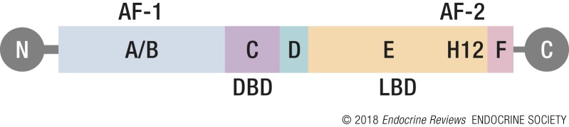
Obligatory domain structure schematic. Drawing showing the six ERα domains, A to F, oriented from the amino (N) to carboxyl (C) terminus. The domains in which each of several key functions is located are indicated: AF-1 and AF-2 mediate transcriptional activity. The DBD interacts with ERE DNA motifs, and the LBD binds E2. Helix 12 (H12) interacts with transcriptional activators and repressors.
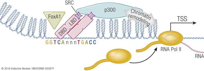
Basic mechanisms of ER-mediated transcriptional regulation. Access to target genes is controlled, in part, by chromatin state. Pioneering factors, such as FOXA1 (green triangle), provide areas of more open chromatin, facilitating access of ER (pink) to ERE DNA motifs. Interaction with E2 leads to recruitment of steroid receptor coactivator (SRC) molecules to ER and interaction with p300. Chromatin remodeling activity of p300, especially histone acetyl transferase activity, facilitates RNA Pol II assembly at the TSS, leading to increased RNA transcription of ER target genes.
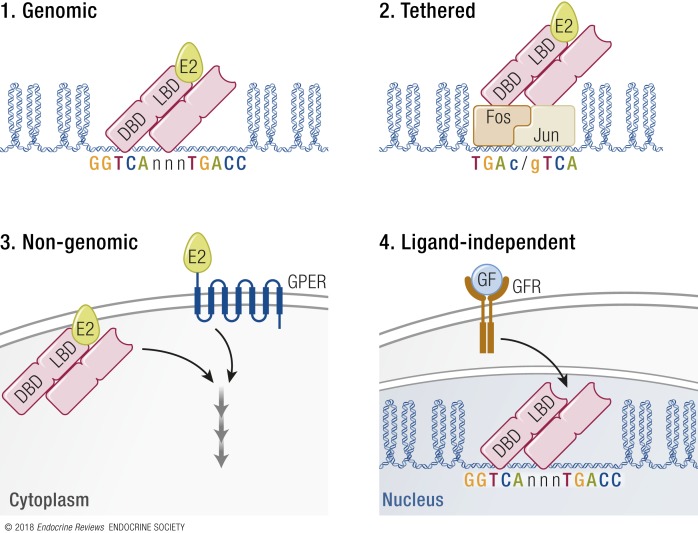
Variations in the basic mechanism of E2 response. Four different E2 response mechanisms have been described. (1) The genomic mechanism involves interaction between ER and ERE DNA motifs. (2) The tethered mechanism involves indirect interaction between ER and other transcriptional regulators, such as the AP-1 DNA motif that binds the FOS/JUN dimer. Thus, ER is “tethered” to the DNA via, in this example, FOS/JUN binding to its AP-1 DNA motif. (3) Nongenomic signaling is so-called because it initiates a signal from extracellular E2 that leads to rapid signal cascades in the cytoplasm, and thus the response does not involve interaction with genomic features. The responses are mediated by membrane-associated ER, or by GPER, a G protein–coupled receptor. (4) Ligand-independent signaling involves transduction of extracellular growth factor (GF) activation of cell membrane GF receptor (GFR), which initiates signaling cascades, such as MAPK. The signal is received by the ER, activating its transcriptional modulation of target genes, despite lacking E2 ligand.
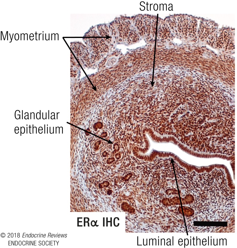
Mouse uterus as ERα-mediated E2 response model. Cross-section of a mouse uterus stained by immunohistochemistry (IHC) with an antibody to ERα illustrating plentiful ERα in all cells, including the luminal and glandular epithelial cells, stromal cells, and myometrial cells. Photo taken with ×10 objective, scale bar = 100 μM.
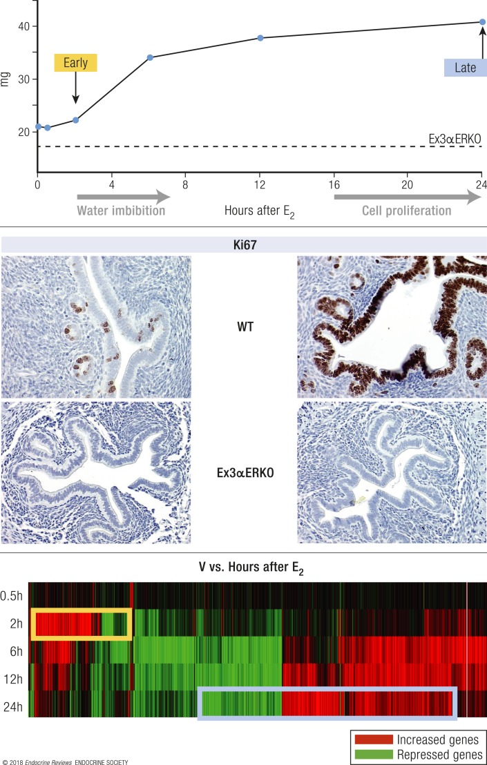
Biphasic uterine response to E2. (Top) Schematic of uterine weight response of WT (solid line) and Ex3αERKO (ERα-null) mice. Administering a single dose of E2 to an ovariectomized female mouse results in an early uterine weight increase, caused by water imbibition, and a later increase due to epithelial cell proliferation. Ex3αERKO mice lack any E2 response. (Middle) Ki67 proliferative marker in samples from WT and Ex3αERKO mice treated for 24 h with vehicle (V, left) or E2 (right). Photos taken with ×20 objective. (Bottom) Heat map of differentially expressed (vs V) transcripts in uterus samples from WT mice treated for 0.5, 2, 6, 12, or 24 h with E2.
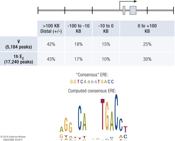
ERα ChIP-seq indicates sites of interaction in mouse uterus. (Top) Schematic and table show positions of ERα-binding peaks relative to a generic gene model. Both pre-bound (V) and E2-induced (1 h E2) peaks are primarily localized distal from genes. (Bottom) Consensus ERE DNA motif and motif computed from actual ERα binding in uterine tissue.
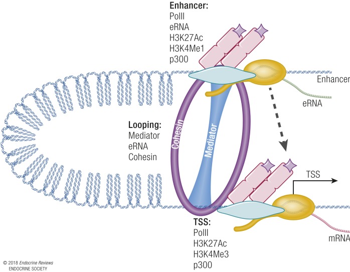
Model of looping to facilitate interaction between enhancers and promoters/TSSs. RNA PolII, enhancer RNA (eRNA) transcription, acetylation of histone H3 lysine 27 (H3K27Ac), monomethylation of histone H3 lysine 4 (H3K4Me1), and p300 are found at enhancers. TSSs have PolII, H3K27Ac, trimethylation of histone H3 lysine 4 (H3K4Me3), and p300. Cohesin/mediator form a looping structure that facilitates interaction between the enhancer and TSSs (dashed arrow).
References
-
- Aranda A, Pascual A. Nuclear hormone receptors and gene expression. Physiol Rev. 2001;81(3):1269–1304. - PubMed
-
- Gibson DA, Saunders PT. Estrogen dependent signaling in reproductive tissues—a role for estrogen receptors and estrogen related receptors. Mol Cell Endocrinol. 2012;348(2):361–372. - PubMed
-
- McEwan IJ. The nuclear receptor superfamily at thirty. Methods Mol Biol. 2016;1443:3–9. - PubMed
Publication types
MeSH terms
Substances
Grants and funding
LinkOut - more resources
Full Text Sources
Other Literature Sources
