. 2018 Aug 15;9(8):2001-2008.
doi: 10.1021/acschemneuro.8b00089. Epub 2018 Jun 20.

Functional Peptidomics: Stimulus- and Time-of-Day-Specific Peptide Release in the Mammalian Circadian Clock

Functional Peptidomics: Stimulus- and Time-of-Day-Specific Peptide Release in the Mammalian Circadian Clock

Norman Atkins Jr et al. ACS Chem Neurosci. .

Abstract

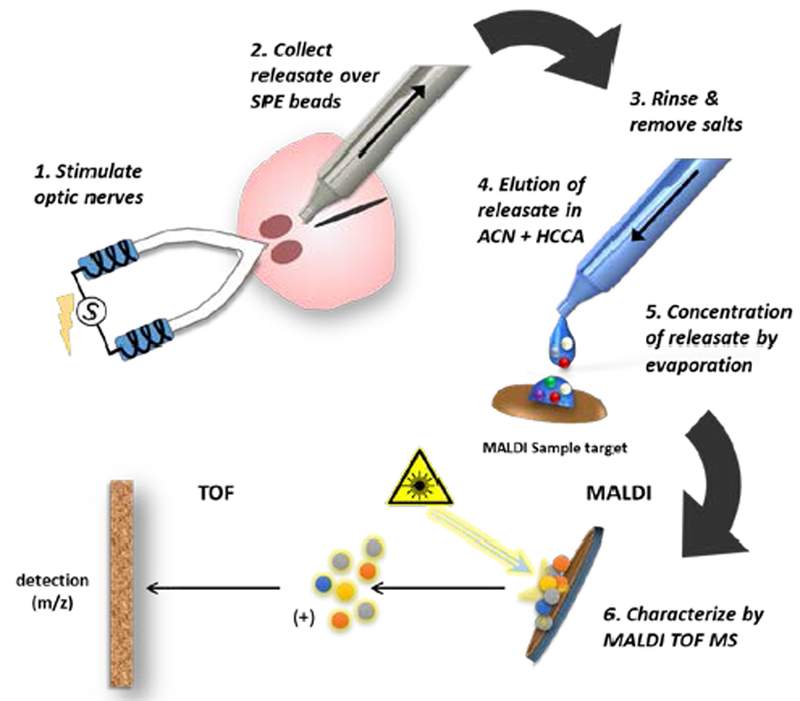
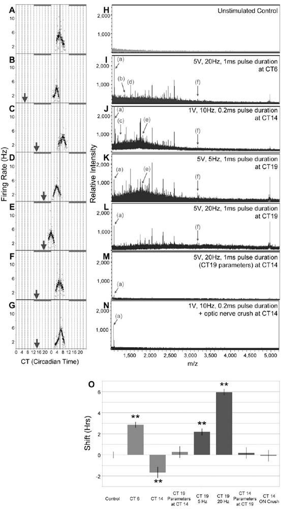
Daily oscillations of brain and body states are under complex temporal modulation by environmental light and the hypothalamic suprachiasmatic nucleus (SCN), the master circadian clock. To better understand mediators of differential temporal modulation, we characterize neuropeptide releasate profiles by nonselective capture of secreted neuropeptides in an optic nerve horizontal SCN brain slice model. Releasates are collected following electrophysiological stimulation of the optic nerve/retinohypothalamic tract under conditions that alter the phase of the SCN activity state. Secreted neuropeptides are identified by intact mass via matrix-assisted laser desorption/ionization time-of-flight mass spectrometry (MALDI-TOF MS). We found time-of-day-specific suites of peptides released downstream of optic nerve stimulation. Peptide release was modified differentially with respect to time-of-day by stimulus parameters and by inhibitors of glutamatergic or PACAPergic neurotransmission. The results suggest that SCN physiology is modulated by differential peptide release of both known and unexpected peptides that communicate time-of-day-specific photic signals via previously unreported neuropeptide signatures.

Keywords: Circadian clock; SCN; mass spectrometry; neuropeptidomics; optic nerve; suprachiasmatic nucleus.


Figures

Figure 1.
Figure 1.
Schematic of the workflow for neuropeptide collection from the SCN brain slice and MS analysis. 1. The optic nerves (ON) attached to a horizontal SCN brain slice are stimulated in tandem by suction electrodes or the SCN is treated directly by droplet containing a chemical stimulus. 2. Releasate is aspirated through a micropipette containing solid-phase extraction beads (SPEs), which bind peptides based on their charge. 3. The beads are rinsed to remove salts and then samples (represented as small colored circles) are transferred to a MALDI target surface. 4. Bound analytes are eluted with acetonitrile (ACN) and addition of a cyano-4 hydroxycinnamic acid (HCCA) MALDI matrix solution. 5. As the acetonitrile evaporates, analytes are concentrated with MALDI matrix onto discrete hydrophobic regions within the HCCA crystals on the pre-treated target plate. 6. The sample is volatized and ionized by matrix-assisted laser desorption/ionization (MALDI). Following ionization, the analytes are subjected to a mass analyzer and detector for spectrophotometric analysis where their mass/charge ratio (m/z) is determined.
Figure 2.
Figure 2.
Stimulus parameters applied to the ON to induce temporal phase shifting and peptide release in the SCN and outcomes. (A) Three different 5-min electrical stimulation paradigms were utilized: Orange, 1 V, 10 Hz, 0.2 ms pulse duration; Green, 5 V, 5 Hz, 1 ms pulse duration; Purple, 5 V, 20 Hz, 1 ms pulse duration. (B) These three stimulation parameters at distinct temporal windows caused different patterns of peptide release (■), and phase shifts of a delay of ~ 2 hr (φD), advance of ~2–3 hr (φA), or an advance of ~ 6 hr (φA+), respectively. N.R., no releasate measured.
Figure 3.
Figure 3.
Stimulus- and time-of-day specific phase-shifts of SCN neuronal activity are accompanied by stimulus- and time-of-day-specific peptide release. Horizontal suprachiasmatic brain slices of the mediobasal hypothalamus and preserving the ONs, which contain retinal afferents to the SCN, were monitored in vitro. (A) The SCN displays a circadian rhythm in neuronal firing rate that peaks at mid-subjective day, CT 6. ONs were stimulated bilaterally via suction electrodes with specific current, frequency, and pulse duration parameters at circadian time points (arrowheads). During daytime (CT 6) and late subjective nighttime (CT 19), ON stimulation evokes phase advance of the neuronal activity rhythm (B, D, and E), while effective parameters at early subjective nighttime (CT 14) trigger phase delay of SCN firing rhythm (C). MALDI-TOF-MS analyses of releasate collected from the SCN exhibit time-of-day and stimulus-specific peptide release profiles (H-L). Some peptide peaks, such as arginine vasopressin (a) and galanin (f), are observed at every timepoint, while release of other peptides, such as neurokinin-B (b), somatostatin-28 (c), angiotensin (d), and little SAAS (e) is restricted to specific time-of-day and stimulus conditions. Effective parameters at CT 19 are ineffective in phase shift (F) and peptide release (M) at CT 14. Transectional ON crush prevents stimulus-evoked phase shifting (G)and peptide release (N). Phase-shifting responses to ON stimulation depend upon unique, time-of-day-effective stimulus parameters (O). **, p < 0.001, one-way ANOVA.

Similar articles

Cited by

References

    1. Hastings MH; Maywood ES; Reddy AB, Two decades of circadian time. J Neuroendocrinol 2008, 20 (6), 812–9. - PubMed
    1. Robinson I; Reddy AB, Molecular mechanisms of the circadian clockwork in mammals. FEBS Lett 2014, 588 (15), 2477–83. - PubMed
    1. Rosenwasser AM; Turek FW, Neurobiology of Circadian Rhythm Regulation. Sleep Med Clin 2015, 10 (4), 403–12. - PubMed
    1. Abrahamson EE; Moore RY, Suprachiasmatic nucleus in the mouse: retinal innervation, intrinsic organization and efferent projections. Brain Res 2001, 916 (1–2), 172–91. - PubMed
    1. Welsh DK; Takahashi JS; Kay SA, Suprachiasmatic nucleus: cell autonomy and network properties. Annu Rev Physiol 2010, 72, 551–77. - PMC - PubMed

Publication types

Substances

LinkOut - more resources