. 2018 Dec;91(1092):20170930.
doi: 10.1259/bjr.20170930. Epub 2018 Jun 29.

Quantitative magnetic resonance imaging and radiogenomic biomarkers for glioma characterisation: a systematic review

Affiliations

Quantitative magnetic resonance imaging and radiogenomic biomarkers for glioma characterisation: a systematic review

Pohchoo Seow et al. Br J Radiol. 2018 Dec.

Abstract

Objective:: The diversity of tumour characteristics among glioma patients, even within same tumour grade, is a big challenge for disease outcome prediction. A possible approach for improved radiological imaging could come from combining information obtained at the molecular level. This review assembles recent evidence highlighting the value of using radiogenomic biomarkers to infer the underlying biology of gliomas and its correlation with imaging features.

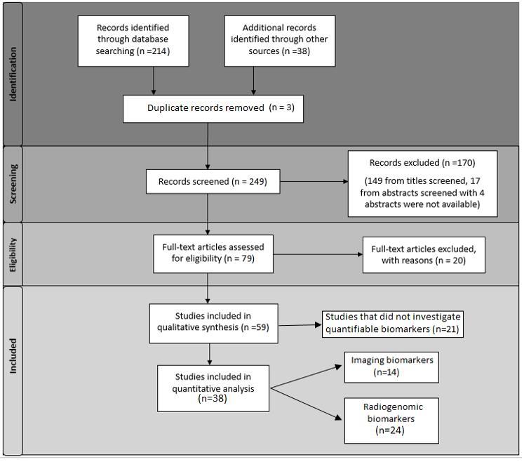
Methods:: A literature search was done for articles published between 2002 and 2017 on Medline electronic databases. Of 249 titles identified, 38 fulfilled the inclusion criteria, with 14 articles related to quantifiable imaging parameters (heterogeneity, vascularity, diffusion, cell density, infiltrations, perfusion, and metabolite changes) and 24 articles relevant to molecular biomarkers linked to imaging.

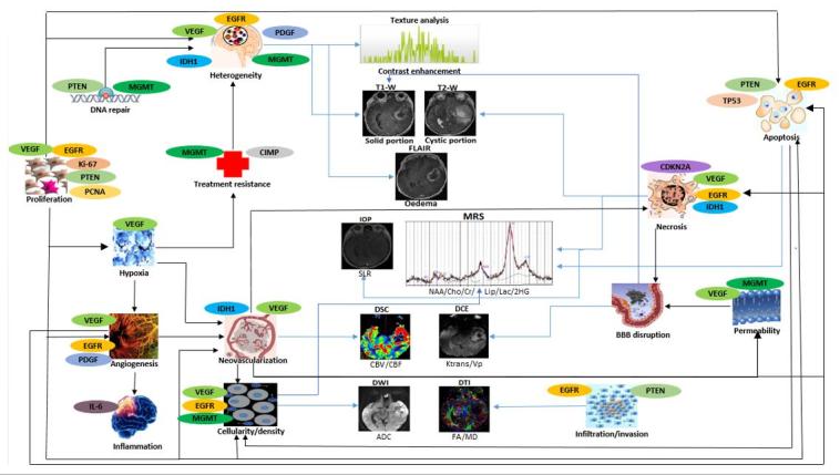
Results:: Genes found to correlate with various imaging phenotypes were EGFR, MGMT, IDH1, VEGF, PDGF, TP53, and Ki-67. EGFR is the most studied gene related to imaging characteristics in the studies reviewed (41.7%), followed by MGMT (20.8%) and IDH1 (16.7%). A summary of the relationship amongst glioma morphology, gene expressions, imaging characteristics, prognosis and therapeutic response are presented.

Conclusion:: The use of radiogenomics can provide insights to understanding tumour biology and the underlying molecular pathways. Certain MRI characteristics that show strong correlations with EGFR, MGMT and IDH1 could be used as imaging biomarkers. Knowing the pathways involved in tumour progression and their associated imaging patterns may assist in diagnosis, prognosis and treatment management, while facilitating personalised medicine.

Advances in knowledge:: Radiogenomics can offer clinicians better insight into diagnosis, prognosis, and prediction of therapeutic responses of glioma.


Figures

Figure 1.
Figure 1.
Literature assessment. Flow diagram of literature assessment.
Figure 2.
Figure 2.
A case of grade IV GBM with EGFR amplification/overexpression. MRI features showing greater ratio of T2 bright volume to the enclosed T1 enhancing volume in GBM: (a) CUBE FLAIR images depicting perilesional oedema, (b) calculated 3D-T2 bright volume (147.62 cm3), (c) T1W post-contrast showing Rt parietal enhancing GBM with internal necrosis, and (d) calculated 3D-T1-enhancing volume including internal necrosis (41.03 cm3). EGFRendothelial growth factor receptor; GBM, glioblastoma multiformes.
Figure 3.
Figure 3.
MRI post-gadolinium images of various grade IV GBM with hypermethylated and unmethylated MGMT. Imaging features showing: (a) mixed-nodular in a patient with hypermethylated MGMT and (b) ring enhancement in unmethylated MGMT. Another two cases demonstrating (c) preferential location of grade IV GBM with hypermethylation of the MGMT promoter located in parietal and occipital lobes, and (d) unmethylated of the MGMT promoter in the temporal lobes. GBM, glioblastoma multiformes; MGMTO6-methylguanine-DNA-methyltransferase.
Figure 4.
Figure 4.
MRI features of various grade IV GBM patients with IDH1 mutation. The features included T2W images showing (a) large size at time of diagnosis (b) multifocality, and (c) cystic components. (d) Post-gadolinium T1W showing non-enhancing solid tumour component, (e) greater frequency of contact with the ventricles, and *(f) usually less necrotic (<50% of tumour volume, and (g) T2 FLAIR showing less perilesional oedema (<50% of tumour volume). GBM, glioblastoma multiformes; IDH1, isocitratedehydrogenase 1.
Figure 5.
Figure 5.
MRI features of grade III ODG patients with of 1p/19q co-deletion. Imaging findings showing: (a) indistinct borders on T1W, (b) GRE sequence with paramagnetic susceptibility and calcification and, (c-d) mixed signal intensities on T1W and T2W. GRE, gradient echo; ODG, oligodendroglioma.
Figure 6.
Figure 6.
The MRI images of a grade IV GBM with prominent palisading necrosis, microvascular proliferation, Ki-67 index ~15–20% in a few cellular areas. Imaging findings showing: (a) relative CBV colour map where high blood volume was seen at the rim area, (b) decreased ADC shown as hypointense area compared to CSF in tumour region, (c) the voxel placement in SVS, and (d) the corresponding brain spectra acquired using LC Model where MI, Cho, Cr, NAA & Lip peaks are labelled. Elevated lipid peaks and Cho, with decreased NAA were apparent in the spectrum. ADC, apparent diffusion co-efficient; CBV, cerebral blood volume; Cho, choline; Cr, creatine; CSF, cerebrospinal fluid; GBM, glioblastoma multiformes; Lip, lipid; MI, myo-inositol; NAA, N-acetyl aspartate; SVS, single voxel spectroscopy.
Figure 7.
Figure 7.
Radiogenomic approach for glioma characterisation. A schematic diagram to illustrate the relationship of glioma morphology with gene expressions and imaging characteristic. Black arrows indicate associations between different glioma morphology while blue arrows represent the linking between glioma morphology and MRI. The images displayed are for visual guide only. ADC, apparent diffusion co-efficient; BBB, blood-brain barrier; CBF, cerebral blood flow;CBV, cerebral blood volume; CDKN2A,cyclin-dependent kinase inhibitor; Cho, choline; Cr, creatine; DCE, dynamic contrast-enhanced; DSC, dynamic susceptibility contrast; DWI, diffusion-weighted imaging; DTI, diffusion tensor imaging; EGFR, endothelial growth factor receptor; FA, fractional anisotropy; IDH1, isocitratedehydrogenase 1; IOP, in and opposed-MRI; Ktrans, volume transfer constant; Lac, lactate; Lip, lipid; MD, mean diffusivity; MGMT,O6-methylguanine-DNA-methyltransferase; MRS, magnetic resonance spectroscopy; NAA, N-acetyl aspartate; PCNA, proliferating cell nuclear antigenPDGFplatelet-derived growth factor; PTENphosphatase andtensin homolog; SLR, signal loss ratio; VEGFvascularendothelial growth factor; VP, plasma volume.

References

    1. Ostrom QT, Gittleman H, de Blank PM, Finlay JL, Gurney JG, McKean-Cowdin R, et al. American brain tumor association adolescent and young adult primary brain and central nervous system tumors diagnosed in the United States in 2008-2012. Neuro Oncol 2016; 18(suppl 1): i1–i50. doi: 10.1093/neuonc/nov297 - DOI - PMC - PubMed
    1. Kleihues P, Louis DN, Scheithauer BW, Rorke LB, Reifenberger G, Burger PC, et al. The WHO classification of tumors of the nervous system. J Neuropathol Exp Neurol 2002; 61: 215–25. doi: 10.1093/jnen/61.3.215 - DOI - PubMed
    1. Louis DN, Perry A, Reifenberger G, von Deimling A, Figarella-Branger D, Cavenee WK, et al. The 2016 World Health Organization classification of tumors of the central Nervous system: a summary. Acta Neuropathol 2016; 131: 803–20. doi: 10.1007/s00401-016-1545-1 - DOI - PubMed
    1. Guzmán-De-Villoria JA, Mateos-Pérez JM, Fernández-García P, Castro E, Desco M, et al. Added value of advanced over conventional magnetic resonance imaging in grading gliomas and other primary brain tumors. Cancer Imaging 2014; 14: 35. doi: 10.1186/s40644-014-0035-8 - DOI - PMC - PubMed
    1. Lambin P, Rios-Velazquez E, Leijenaar R, Carvalho S, van Stiphout RGPM, Granton P, et al. Radiomics: Extracting more information from medical images using advanced feature analysis. Eur J Cancer 2012; 48: 441–6. doi: 10.1016/j.ejca.2011.11.036 - DOI - PMC - PubMed

Publication types