

The hepatic pre-metastatic niche in pancreatic ductal adenocarcinoma
- PMID: 29903049
- PMCID: PMC6003100
- DOI: 10.1186/s12943-018-0842-9
The hepatic pre-metastatic niche in pancreatic ductal adenocarcinoma
Abstract
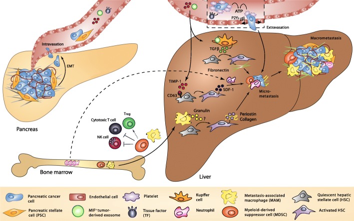
Pancreatic ductal adenocarcinoma (PDAC) remains one of the most aggressive malignancies to date, largely because it is associated with high metastatic risk. Pancreatic tumors have a characteristic tendency to metastasize preferentially to the liver. Over the past two decades, it has become evident that the otherwise hostile milieu of the liver is selectively preconditioned at an early stage to render it more conducive to the engraftment and growth of disseminated cancer cells, a concept defined as pre-metastatic niche (PMN) formation. Pancreatic cancer cells exploit components of the tumor microenvironment to facilitate their migration out of the primary tumor, which often involves conversion of pancreatic cancer cells from an epithelial to a mesenchymal phenotype via the epithelial-to-mesenchymal transition. Pancreatic stellate cells and matrix stiffness have been put forward as major drivers of invasiveness in PDAC. Even before the onset of pancreatic cancer cell dissemination, soluble factors and extracellular vesicles secreted by the primary tumor, and possibly even premalignant lesions, help shape a supportive niche in the liver by providing vascular docking sites for circulating tumor cells, enhancing vascular permeability, remodeling the extracellular matrix and recruiting immunosuppressive inflammatory cells. Emerging evidence suggests that some of these tumor-derived factors may represent powerful diagnostic or prognostic biomarkers. Though our understanding of the mechanisms driving PMN formation in PDAC has expanded considerably, many outstanding questions and challenges remain. Further studies dissecting the molecular and cellular events involved in hepatic PMN formation in PDAC will likely improve diagnosis and open new avenues from a therapeutic standpoint.
Keywords: Metastasis; Niche; Pancreatic cancer; Stroma.
Conflict of interest statement
Ethics approval and consent to participate
Not applicable.
Competing interests
The authors declare that they have no competing interests.
Publisher’s Note
Springer Nature remains neutral with regard to jurisdictional claims in published maps and institutional affiliations.
Figures
References
-
- Åkerberg D, Ansari D, Andersson R, Tingstedt B. The effects of surgical exploration on survival of unresectable pancreatic carcinoma: a retrospective case-control study. J Biomed Sci Eng. 2017;10:1–9. doi: 10.4236/jbise.2017.101001. - DOI
Publication types
MeSH terms
LinkOut - more resources
Full Text Sources
Other Literature Sources
Medical
Miscellaneous
