Review
. 2018:154:129-149.
doi: 10.1016/B978-0-444-63956-1.00008-4.

The neuropathology of the adult cerebellum

Affiliations
Review

The neuropathology of the adult cerebellum

Arnulf H Koeppen. Handb Clin Neurol. 2018.

Abstract

This chapter summarizes the neuropathologic features of nonneoplastic disorders of the adult cerebellum. Gait ataxia and extremity dysmetria are clinical manifestations of diseases that interrupt the complex cerebellar circuitry between the neurons of the cerebellar cortex, the cerebellar nuclei (especially the dentate nuclei), and the inferior olivary nuclei. The cerebellum is a prominent target of several sporadic and hereditary neurodegenerative diseases, including multiple system atrophy, spinocerebellar ataxia, and Friedreich ataxia. Purkinje cells display selective vulnerability to hypoxia but a surprising resistance to hypoglycemia. A classic toxin that damages the cerebellar cortex is methylmercury, but the most common injurious agent to Purkinje cells is ethanol. Many drugs cause ataxia, but doubts continue about phenytoin. Ischemic lesions of the cerebellum due to arterial thrombosis or embolism cause a spectrum of symptoms and signs, depending on the territory involved. Large hemorrhages have an unfavorable prognosis because they displace critical brainstem structures or penetrate into the fourth ventricle. Fungal infections and toxoplasmosis of the cerebellum, and cerebellar progressive multifocal leukoencephalopathy, have become rarer because of improved control of the acquired immunodeficiency syndrome. Ataxia is a prominent feature of prion disease. Adult-onset Niemann-Pick type C1 disease and Kufs disease may have a predominantly ataxic clinical phenotype. The adult cerebellum is also vulnerable to several leukodystrophies. A rare but widely recognized complication of cancer is paraneoplastic cerebellar degeneration.

Keywords: Friedreich ataxia; cerebellar circuitry; hypoglycemia; hypoxia; infections; leukodystrophy; lipidoses; multiple sclerosis; multiple system atrophy; paraneoplastic cerebellar degeneration; spinocerebellar ataxia; stroke; toxicity.


Figures

Fig. 8.1.
Fig. 8.1.
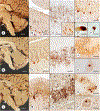
The neurons of the reciprocal cerebellar circuitry. (AD) Cerebellar cortex; (E and F) dentate nuclei (DN); (G and H) inferior olivary nuclei (ION). Immunohistochemical stains: (A) class III b-tubulin; (B, F, and H) glutamic acid decarboxylase (GAD); (C) vesicular glutamate transporter 2 (VGluT2); (D) vesicular glutamate transporter 1 (VGluT1); (E), nonphosphorylated neurofilament protein; and (G) calbindin D28k. (A) The Purkinje cells of the cerebellar cortex show their well-known idiodendritic tree. Small neurons in the molecular layer that also react with the antibody to class III b-tubulin represent basket and stellate cells. In the granular layer, a Golgi neuron is immunoreactive (arrow). (B) The GAD stain of the cerebellar cortex shows pale reaction product in the cytoplasm of Purkinje cells and more robust reaction product in terminal baskets. Anti-GAD also labels parallel fibers and basket and stellate cells in the molecular layer, and the axonal plexuses of Golgi neurons in the granular layer (arrow). (C) An antibody to VGluT2 generates punctate reaction product in climbing fibers (arrow) as they abut Purkinje cell dendrites, and more intense labeling of mossy fiber terminals in the granular layer. (D) Anti-VGluT1 labels mossy fiber terminals in the granular layer. Strong, finely granular, reaction product is distributed throughout the entire thickness of the molecular layer, generating negative images of primary and secondary dendritic branches of Purkinje cells. (E) The neurofilament stain shows reaction product in large, intermediate, and small neurons of the dentate nucleus. (F) Anti-GAD generates reaction product in axon terminals of the DN and creates negative images of larger neurons (N). The stain also visualizes GAD in the cytoplasm of small neurons at the perimeter of the DN (arrows). (G) Neuronal cell bodies and dendrites of the ION are strongly reactive with anticalbindin D28k. (H) Terminals abutting ION neurons are strongly GAD-reactive. Bars, 50 μm.
Fig. 8.2.
Fig. 8.2.
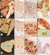
The cerebellar and olivary lesions in multiple system atrophy (MSA), spinocerebellar ataxia types 1 (SCA-1) and 2 (SCA-2). (AF) MSA; (GK) SCA-1; (LP) SCA-2. Immunohistochemical stains: (B, H, and M) cerebellar cortex, class III b-tubulin; (C I, and N) dentate nuclei (DN), glutamic acid decarboxylase (GAD); (C, inset) DN, neuron-specific enolase; (D, J, and O) inferior olivary nuclei (ION), calbindin D28k. (E) Neuron in the basis pontis, ubiquitin; (F) neuron in the basis pontis, a-synuclein; (K and P) neurons in the basis pontis, 1C2 for polyglutamine. The midline sections of the brains in MSA (A), SCA-1 (G), and SCA-2 (L) show small cerebella and, in MSA (A) and SCA-2 (L), atrophy of the basis pontis (arrows). The cerebellar cortex dis-plays thinning of the molecular layer, loss of Purkinje cells, and an impoverished dendritic tree. The DN shows low density of GAD-reactive terminals in MSA (C), though DN neurons are intact (C, inset). In SCA-1 (I) and SCA-2 (N), the DN displays a greater density of GAD-reactive terminals than in MSA (C). Void spaces corresponding to large DN neurons are absent in MSA (C), SCA-1 (I), and SCA-2 (N). The ION in MSA (D), SCA-1 (J), and SCA-2 (O) reveal gaps. Shared features of MSA, SCA-1, and SCA-2 are Purkinje cell atrophy, loss of GABAergic afferents of the DN, and retrograde degeneration of the ION. MSA and SCA-2 share atrophy of the basis pontis. 1C2-positive intranuclear neuronal inclusion bodies indicate poly-glutamine expansions in SCA-1 (K) and SCA-2 (P). Bars: (A, G, and L) 1 cm; (BD C, inset, HJ, and MO, 100 μm; E and F, K, and P, 10 μm (oil immersion).
Fig. 8.3.
Fig. 8.3.
The cerebellar and olivary lesions in spinocerebellar ataxia types 6 (SCA-6) and 3 (SCA-3), and Friedreich ataxia (FA). (AE) SCA-6; (FJ) SCA-3; (KN) FA. Immunohistochemical stains: (B G, and L) cerebellar cortex, class III b-tubulin; (C, H, and M) dentate nuclei (DN), glutamic acid decarboxylase (GAD); (D, I, and N) inferior olivary nuclei (ION), D28k; (E and J) neurons of the basis pontis, 1C2 for polyglutamine. The midline section of the brain in SCA-6 (A) shows atrophy of the upper vermis with wide spaces between folia. The pons is well preserved in SCA-6 (A). Pontine atrophy, however, is present in the illus-trated case of SCA-3 (F, arrow). Collapse of the DN in FA is especially apparent on a macroscopic stain for iron (K). The cerebellar cortex in SCA-6 (B) shows loss of Purkinje cells whereas the cortex in SCA-3 (G) and FA (L) is normal. The DN in SCA-6 shows reduced density of GABAergic terminals (C). The DN in SCA-3 (H) and FA (M) displays grumose reaction (arrows). Voids representing large neurons are still visible in the DN of SCA-3 (H). The ION in SCA-6 (D) reveals minor gaps in the chain of neurons, but the spherical dendritic tree in the remaining nerve cells is intact. The ION is entirely normal in SCA-3 (I) and FA (N), reflecting the integrity of Purkinje cells (G and L). 1C2-positive neuronal inclusion bodies are cytoplasmic in SCA-6 (E, arrows) but intranuclear in SCA-3 (J, arrow). Bars: (A, F, and K) 1 cm; (BD, GI, and LN) 100 μm; (E and J), 10 μm (oil immersion).
Fig. 8.4.
Fig. 8.4.
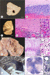
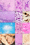
The cerebellum in hypoxia. (A, B) Three-day survival after incomplete cardiorespiratory resuscitation (49-year-old man); (C, D) 6-day survival after a 20-minute-long episode of pulselessness and resuscitation (59-year-old man); (EG) 3-week survival after near drowning (14-year-old girl); (H, I) 1-year-survival after unsuccessful suicide attempt by hanging (48-year-old man). Stains: (A, C, F, G, and I) hematoxylin and eosin stains of cerebellar cortex; (B and D), hematoxylin and eosin stain of the DN; (D, inset) IBA-1 immunostain for microglia in the dentate nuclei (DN). (E and H) gross specimens. Brief stagnant hypoxia causes loss of basophilic staining of Purkinje cells (A, and inset) and DN neurons (B). The granular layer is intact. After longer survival, loss of Purkinje cells is complete (C). Neurons of the DN still show the acidophilia of hypoxia (D), but a microglial response is now also apparent (D, inset). In the case of near drowning (EG), the cerebellar cortex becomes atrophic, and the white matter of the brachium pontis is peculiarly white. Sections show thinning of the molecular layer and total loss of Purkinje and granule cells (F). The Purkinje cell layer displays intense proliferation of Golgi epithelial cells (“Bergmann gliosis”) (G). The cerebellum in the case of stagnant hypoxia during an unsuccessful suicide attempt by hanging (H) shows gross cerebellar atrophy. Loss of Purkinje cells is complete, and the density of granule cells is reduced (I). Bars: (A, C, F, and I) 100 μm; (B and D) 50 μm; (A and D, insets, and G) 20 μm; (E and H) 1 cm.
Fig. 8.5.
Fig. 8.5.
The cerebellum in hypoglycemia. (A) Parietal cortex; (B) cerebellar cortex. A forensic autopsy was conducted on a 27-year-old man who was found in an unexplained comatose state from which he did not recover over a period of 3 weeks. Blood glucose levels were low, and the diagnosis was accidental, suicidal, or homicidal injection of insulin. The deeper layers of the parietal cortex (A) show necrosis, proliferation of capillaries, and infiltration by macrophages. The subpial gray matter (to the upper right) displays reactive astrogliosis (A). In contrast to the cerebral cortex, the cerebellar cortex is intact (B). Stain: hematoxylin and eosin; bars: 50 μm.
Fig. 8.6.
Fig. 8.6.
The cerebellum in ethanol and mercury toxicity. (AC) Alcoholic cerebellar degeneration; (D and E) cerebellar atrophy in methylmercury toxicity. The gross specimen of the cerebellum obtained from a 58-year-old man with alcoholic cerebellar degeneration reveals atrophy of the superior vermis (A). Stains: (B, D, E, and E, inset), hematoxylin and eosin; (C and C, inset) Bodian silver stain for axons; (D, inset) immunohistochemistry of phosphorylated neurofilament proteins. Sections show loss of Purkinje cells (B), empty baskets (C, arrow) and axonal expansions (torpedoes) in the granular layer (C, inset). The section of the cerebellum in chronic methylmercury toxicity shows depletion of granular cells immediately adjacent to the Purkinje cell layer (D, arrow). Basket fibers surround intact Purkinje cells or form empty baskets (D, inset). The dentate nucleus displays moderate neuronal loss (E) and regions of grumose regeneration (E, inset). Bars: (A) 1 cm; (B) 200 μm; (D and E) 50 μm; (CE, insets) 20 μm. (D and E courtesy of Professor F. Ikuta, Niigata, Japan).
Fig. 8.7.
Fig. 8.7.
Vascular lesions of the cerebellum. (A and B) Small spontaneous cerebellar hemorrhage in a 68-year-old man with cancer; (CE) 7-day-old hemorrhagic infarction in a 71-year-old man with cerebral embolism arising from vegetations on the aortic valve; (FH) perioperative thrombosis of the right superior cerebellar artery in a 68-year-old man (survival of 2 months). (IL), small infarction of the dentate nucleus (DN) (I, arrow) and olivary hypertrophy (J, arrow) in an 88-year-old man. Stains: (B, D, E, G, and H) hematoxylin and eosin; (I) Weil stain for myelin; (JL) Bodian silver stain. The acute cerebellar hemorrhage (A) displaces the cerebellar cortex, but has not yet generated a cellular response (B). In contrast, the hemorrhagic infarction (C) causes hypoxia in Purkinje cells (D), extravasation of red blood cells (D, arrow), and a vigorous macrophage response (E) with erythrophagocytosis (E, arrow). The old cystic infarction (F) is due to thrombotic occlusion of the superior cerebellar artery (F, arrow). The infarction involves the full thickness of the cerebellar cortex though Bergmann glia and their parent cell bodies remain intact (G). The infarction is sharply demarcated from the intact tissue (G). Small macrophages are present in the cystic infarction (H), and some contain brown hemosiderin pigment. A small infarction of the DN (I, arrow) causes transsy-naptic hypertrophy of the inferior olivary nucleus (J, arrow), characterized by nerve cell vacuolation (K) and a bizarre plaque-like dendritic expansion (L). Bars, (A, C, and I) 1 cm; (F) 2 cm; (J) 5 mm; (B, D, and G) 100 μm; (E, H, K, and L) 20 μm.
Fig. 8.8.
Fig. 8.8.
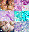
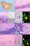
The cerebellum in leptomeningitis. (AD) Streptococcus pneumoniae; (EG) Aspergillus sp.; (HJ) Mycobacterium tuberculosis. (A) and (B) An inflammatory infiltrate fills the subarachnoid space over the cerebellar cortex of this 61-year-old man with S. pneumoniae leptomeningitis. On the gross specimen, the exudate is visible in perivascular spaces (A, arrow). Inflam-matory cells are predominantly polymorphonuclear leukocytes (C). On a smear preparation of pelleted cerebrospinal fluid, the organisms occur as “cocci-inpairs,” are surrounded by a capsule, and are mostly extracellular (D). In the case of incidental Aspergillus sp. leptomeningitis in a 65-year-old man with steroid-induced immunosuppression (EG), the subarachnoid exudate consists of lymphocytes and histiocytes (E). The vessel wall is also infiltrated (E). A Grocott methenamine silver stain shows abundant septate fungal hyphae (F) that display branching (G). (H) A thick exudate is present over brainstem and cerebellum of a 37-year-old woman with thalamic tuberculoma and tuberculous leptomeningitis. Note also the presence of tonsillar herniation. The exudate consists of epithelioid cells, lymphocytes, and Langhans multinucleated giant cells (I). Caseation is absent. A section stained for acid-fast bacilli shows a single red organism (J, arrow). Stains: (B, C, E, and I) hematoxylin and eosin; (D) Gram stain with safranin counterstain; (F, and G) Grocott methenamine silver stain for fungi; (J) Ziehl–Neelsen stain for acid-fast bacilli. Bars: (A and H) 1 cm; (B) 0.5 mm; (E, F, and I) 50 μm; (C and G) 20 μm; (D and J) 10 μm (oil immersion).
Fig. 8.9.
Fig. 8.9.
Infections of the cerebellar parenchyma. (AC) Cerebellitis and Toxoplasma gondii infection in acquired immunodeficiency syndrome (AIDS)/human immunodeficiency virus (HIV); (DG) cytomegalovirus infection in AIDS/HIV; (HJ) progressive multifocal leukoencephalopathy in non-AIDS immunosuppression; (KM) prion disease: sporadic Jakob–Creutzfeldt disease. (A) The cerebellar cortex of a 50-year-old man with AIDS/HIV shows microglial nodules in the molecular layer (A, arrows). (B) Multiple cysts of T. gondii are present in the dentate nucleus (DN). (C) This microphotograph shows an enlarge-ment of four cysts in (B) that lie adjacent to a DN neuron (arrow). (D) The cerebellar section of a 49-year-old man with AIDS/HIV shows a necrotizing disease process in the molecular and granular layers of the cerebellum with many large cells due to cytomeg-alovirus infection. (E) The necrotic cerebellar white matter in this case displays a large cell of unknown derivation with densely amphophilic cytoplasm and a Cowdry type A intranuclear inclusion (arrow). (F and G) These microphotographs show immuno-histochemical reaction product of cytomegalovirus-specific cytoplasmic protein in the molecular (F) and granular layers (G). (HJ) The cerebellar white matter of a 76-year-old man with progressive multifocal leukoencephalopathy due to immunosuppression in the course of chemotherapy for leukemia shows a focus of myelin loss (H), inflammatory infiltration by plasma cells and lymphocytes (I), and a giant astrocyte (I, arrow). (J) The nearby intact white matter displays a pan-nuclear inclusion body (arrow). (K) The gross specimen of a 70-year-old with a rapidly progressive fatal neurologic illness diagnosed as Jakob–Creutzfeldt disease does not show convincing atrophy of the cerebellum. Sections reveal sponginess of the molecular layer (L) and immunohisto-chemical reaction product of protease-resistant prion protein, termed PrPsc in analogy to scrapie (monoclonal antibody 3F4) (M). Bars: (A, L, and M) 50 μm; (D and H) 100 μm; (B, E, F, G, and I) 20 μm; (C and J) 10 μm (oil immersion).
Fig. 8.10.
Fig. 8.10.
Multiple sclerosis (MS) in the cerebellum. (A) A transverse slice through brainstem and cerebellum reveals plaques of MS in the cerebellar white matter and the brachium pontis (arrows). Stains: (B and C) Luxol fast blue–periodic acid–Schiff (LFB-PAS) for myelin; (D) immunohistochemistry (IHC) for myelin basic protein (MBP); (E) IHC for phosphorylated neurofilament protein; (F) IHC for IBA1; (G) IHC for glial fibrillary acidic protein (GFAP). (B and C) The section stained with LFB-PAS reveals a relatively sharp transition from intact white matter to the plaque of MS. (DG) The plaque is located in the upper right portion of the images. (D) A stain for MBP is similar to (C), showing abrupt ending of myelination. (E) Axons traverse the plaque, but their density is lower than normal, and their caliber is small. (F) The junction between plaque and normal white matter displays micro-glial proliferation. (G) At the junction between plaque and normal white matter, a GFAP stain reveals reactive astrocytes. Bars: (A) 1 cm; (B) 0.5 mm; (CG) 50 μm.
Fig. 8.11.
Fig. 8.11.
The cerebellum in adult-onset lipid storage disease. (AI) Niemann–Pick disease type C1; (JM) ceroid-lipofuscinosis (Kufs disease). Stains: (B, C, GJ) hematoxylin and eosin; (D, E, and K) Bodian silver stain; (F, L, M) autofluorescence. (AF) An archival case of a 54-year-old man shows reduced bulk of the dentate nucleus; loss of Purkinje cells (B) and a torpedo (D); lipid storage in Golgi neurons (B and C, arrows); neurofibrillary tangles (E); and autofluorescence of lipid-laden neurons of the granular layer (F). (GI) A genetically confirmed recent case of a 37-year-old woman with progressive ataxia shows loss of Purkinje cells (G), lipid storage in a Lugaro cell (G, arrow), a Golgi neuron (H), and a dentate nucleus neuron (I). (JM). The cerebellar cortex in Kufs disease reveals reduced numbers of Purkinje cells and neurons in the granular layer (J). Some Purkinje cells show unusual dendritic expansions (K). The Purkinje cell cytoplasm is autofluorescent (L and M). Bars: (B, D, G, and K) 50 μm; (C, E, F, H, I, and M) 20 μm; (J and L) 100 μm.
Fig. 8.12.
Fig. 8.12.
The cerebellum in adultonset autosomal-dominant leukodystrophy. Immunohistochemical stains: (A, B) Myelin basic protein; (C) class III b-tubulin; (D) phosphorylated neurofilament protein; (E) hematoxylin and eosin; (F) IBA1 as a marker of microglia; (G) glial fibrillary acidic protein. (A) The subcortical white matter of the cerebellum in this 54-year-old man shows patchy loss of myelin. (B) In contrast to multiple sclerosis, the demarcation of depleted and intact white matter is not sharp, and axons at the transition zone display bulbous endings (C, D). (C) The stain for class III b-tubulin does not reveal axons passing into the demyelinated region. (D) In contrast to the tubulin stain, reaction product of phosphorylated neurofilament protein reveals delicate axons in the myelindeficient area. (E) Despite extensive myelin loss, oligodendroglia persist. (F) Microglia are abundant in transition zones. (G) The depleted regions show large and bizarre astrocytes while fibrous gliosis is absent. Bars: (A) 0.5 mm; (BG) 50 μm.

References

    1. Alturkustani M, Sharma M, Hammond R et al. (2013). Adult-onset leukodystrophy: review of 3 clinicopathologic phe-notypes and a proposed classification. J Neuropathol Exp Neurol 72: 1090–1103. - PubMed
    1. Ambrosi G, Flace P, Lorusso L et al. (2007). Non-traditional large neurons in the granular layer of the cerebellar cortex. Eur J Histochem 51 (Suppl 1): 59–64. - PubMed
    1. Antinori A, Larussa D, Cingolani A et al. (2004). Prevalence, associated factors, and prognostic determinants of AIDS-related toxoplasmic encephalitis in the era of advanced highly active antiretroviral therapy. Clin Infect Dis 39: 1681–1691. - PubMed
    1. Arsov T, Smith KR, Damiano J et al. (2011). Kufs disease, the major adult form of neuronal ceroid lipofuscinosis, caused by mutations in CLN6. Am J Hum Genet 88: 566–573. - PMC - PubMed
    1. Auer RN, Benveniste H (1997). Hypoxia and related conditions. In: Graham DI, Lantos PL (Eds.), Greenfield’s neuropathology Oxford University Press, New York, pp. 263–314.