. 2018 Jun:32:9-20.
doi: 10.1016/j.ebiom.2018.05.035. Epub 2018 Jun 11.

Oxysterol Signatures Distinguish Age-Related Macular Degeneration from Physiologic Aging

Affiliations

Oxysterol Signatures Distinguish Age-Related Macular Degeneration from Physiologic Aging

Jonathan B Lin et al. EBioMedicine. 2018 Jun.

Abstract

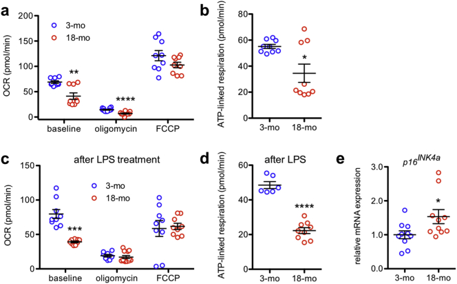
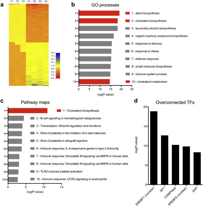
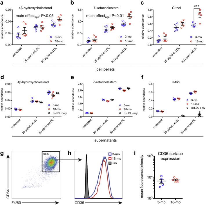
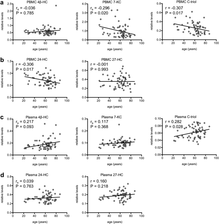
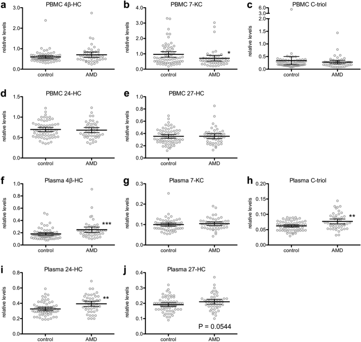
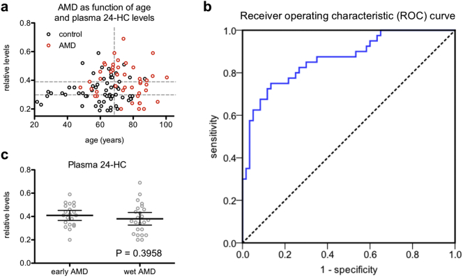
Macrophage aging is pathogenic in numerous diseases, including age-related macular degeneration (AMD), a leading cause of blindness in older adults. Although prior studies have explored the functional consequences of macrophage aging, less is known about its cellular basis or what defines the transition from physiologic aging to disease. Here, we show that despite their frequent self-renewal, macrophages from old mice exhibited numerous signs of aging, such as impaired oxidative respiration. Transcriptomic profiling of aged murine macrophages revealed dysregulation of diverse cellular pathways, especially in cholesterol homeostasis, that manifested in altered oxysterol signatures. Although the levels of numerous oxysterols in human peripheral blood mononuclear cells and plasma exhibited age-associated changes, plasma 24-hydroxycholesterol levels were specifically associated with AMD. These novel findings demonstrate that oxysterol levels can discriminate disease from physiologic aging. Furthermore, modulation of cholesterol homeostasis may be a novel strategy for treating age-associated diseases in which macrophage aging is pathogenic.

Keywords: Age-related macular degeneration; Aging; Cholesterol; Lipids.


Figures

Fig. 1
Fig. 1
Peritoneal macrophages from old mice exhibit quantifiable signs of aging. (a) Aged peritoneal macrophages had reduced oxygen consumption rate (OCR) both at baseline (N = 9/group; 2-tailed, unpaired Welch's t-test) and in response to oligomycin (N = 11–12/group; 2-tailed, unpaired t-test) and significantly reduced ATP-linked respiration (b; N = 9/group; 2-tailed, unpaired Welch's t-test). (c) Lipopolysaccharide (LPS)-treated aged peritoneal macrophages also had impaired mitochondrial bioenergetics at baseline (N = 8–12/group; 2-tailed, unpaired Welch's t-test) and significantly reduced ATP-linked respiration (d; N = 6–9/group; 2-tailed, unpaired t-test) compared to LPS-treated young peritoneal macrophages. (e) Aged peritoneal macrophages had increased mRNA expression of the senescence marker p16INK4a (N = 10/group; 2-tailed, unpaired t-test). Open circles depict individual data points; horizontal lines depict mean ± SEM (a-e) (* P < .05; ** P < .01; *** P < .001; **** P < .0001).
Fig. 2
Fig. 2
Transcriptomic profiling of aged peritoneal macrophages. (a) Aged peritoneal macrophages display numerous transcriptomic changes, which suggest perturbations in various gene ontology (GO) processes (b) and pathway maps (c). (d) Interactome analysis revealed numerous overconnected transcription factors (TFs) whose known gene targets were overrepresented in the genes we identified as dysregulated in aged versus young macrophages.
Fig. 3
Fig. 3
Aged peritoneal macrophages have abnormal oxysterol content. (a-c) Aged peritoneal macrophages contained significantly more intracellular 4β-hydroxycholesterol (4β-HC) and 7-ketocholesterol (7-KC) than their young counterparts at baseline and after treatment with 25 or 50 μg/ml oxidized LDL (oxLDL) (N = 5/group; 2-way ANOVA) and significantly more intracellular cholestane-3β,5α,6β-triol (C-triol) after treatment with 50 μg/ml oxLDL (N = 5/group; 2-way ANOVA with Bonferroni post-hoc test). (d-f) Although some comparisons were statistically significant due to low within-group variance, the supernatants of young and aged peritoneal macrophages contained qualitatively similar levels of 4β-HC, 7-KC, and C-triol both at baseline and after treatment with oxLDL (N = 5/group; 2-way ANOVA with Bonferroni post-hoc test). (g) Representative flow cytometry plot from young and aged peritoneal macrophages showing gating on macrophage markers CD64 and F4/80. (h-i) Young and aged peritoneal macrophages exhibited similar CD36 surface expression. Isotype staining (iso) was identical between groups (N = 5/group; 2-tailed, unpaired t-test). Open circles depict individual data points; horizontal lines depict mean ± SEM (a-f, i) (*** P < .001).
Fig. 4
Fig. 4
Age affects human peripheral blood mononuclear cell (PBMC) and plasma oxysterol signatures. (a-b) There was a significant negative correlation between age and PBMC 7-KC levels, PBMC C-triol levels, and PBMC 24-HC levels in healthy human subjects. (c-d) There was a significant positive correlation between age and plasma C-triol levels and a trend towards a positive correlation between age and plasma 4β-HC levels. Open circles depict individual data points; lines depict the best-fitting linear regression line (a-d; r = Pearson product-moment correlation coefficient; rs = Spearman rank-order correlation coefficient).
Fig. 5
Fig. 5
Age-related macular degeneration (AMD) patients have altered peripheral blood mononuclear cell (PBMC) and plasma oxysterol signatures. (a-j) We measured PBMC and plasma levels of 4β-HC, 7-KC, C-triol, 24-HC, and 27-HC. AMD patients (N = 44–45) had decreased PBMC 7-KC levels (b; 2-tailed Mann-Whitney U test), elevated plasma 4β-HC levels (f; 2-tailed Mann-Whitney U test), elevated plasma C-triol levels (h; 2-tailed Mann-Whitney U test), elevated plasma 24-HC levels (i; 2-tailed Mann-Whitney U test), and a trend towards elevated plasma 27-HC levels (j; 2-tailed, unpaired t-test) compared to non-AMD controls (N = 61). Open circles depict individual data points; horizontal lines depict mean ± 95% confidence intervals (a-j) (* P < .05; ** P < .01; *** P < .001).
Fig. 6
Fig. 6
Plasma 24-HC levels discriminate age-related macular degeneration (AMD) from physiologic aging. (a) We divided patients into tertiles by plasma 24-HC and by age (i.e., above versus below median age) and found that individuals in the highest tertile of plasma 24-HC who were also above median age (top right) had the highest AMD prevalence. Horizontal dashed grey lines demarcate plasma 24-HC tertiles; the vertical dashed grey line indicates the median age. (b) Receiver operating characteristic (ROC) curve showing plasma 24-HC discriminates between AMD patients and control subjects. (c) We did not observe a statistically significant difference in plasma 24-HC levels in early AMD patients versus advanced neovascular (wet) AMD patients (N = 21 early AMD; 24 wet AMD; 2-tailed, unpaired t-test). Open circles depict individual data points (a, c); horizontal lines depict mean ± 95% confidence intervals (c).

Comment in

References

    1. van Leeuwen R., Klaver C.C., Vingerling J.R., Hofman A., de Jong P.T. Epidemiology of age-related maculopathy: a review. Eur J Epidemiol. 2003;18(9):845–854. - PubMed
    1. Ferris F.L., 3rd, Fine S.L., Hyman L. Age-related macular degeneration and blindness due to neovascular maculopathy. Arch Ophthalmol. 1984;102(11):1640–1642. Chicago, Ill: 1960. - PubMed
    1. Sarwar S., Clearfield E., Soliman M.K. Aflibercept for neovascular age-related macular degeneration. Cochrane Database Syst Rev. 2016;2 - PMC - PubMed
    1. Kim J.H., Lee D.W., Chang Y.S., Kim J.W., Kim C.G. Twelve-month outcomes of treatment using ranibizumab or aflibercept for neovascular age-related macular degeneration: a comparative study. Graefe's archive for clinical and experimental ophthalmology = Albrecht von Graefes Archiv fur klinische und experimentelle. Fortschr Ophthalmol. 2016;254(11):2101–2109. - PubMed
    1. Inoue M., Yamane S., Sato S., Sakamaki K., Arakawa A., Kadonosono K. Comparison of time to retreatment and visual function between Ranibizumab and Aflibercept in age-related macular degeneration. Am J Ophthalmol. 2016;169:95–103. - PubMed