. 2018 Sep;8(9):1096-1111.
doi: 10.1158/2159-8290.CD-18-0275. Epub 2018 Jun 14.

Real-time Genomic Characterization of Advanced Pancreatic Cancer to Enable Precision Medicine

Andrew J Aguirre #  1   2   3   4 Jonathan A Nowak #  5   4   6 Nicholas D Camarda #  5   2   7   8 Richard A Moffitt #  9 Arezou A Ghazani  5   2   7 Mehlika Hazar-Rethinam  10 Srivatsan Raghavan  5   2   3   4 Jaegil Kim  2 Lauren K Brais  5 Dorisanne Ragon  5 Marisa W Welch  5 Emma Reilly  5 Devin McCabe  5   2   7   8 Lori Marini  5   6   7 Kristin Anderka  2 Karla Helvie  5   7 Nelly Oliver  5   7 Ana Babic  5   4 Annacarolina Da Silva  5   4   6 Brandon Nadres  10 Emily E Van Seventer  10 Heather A Shahzade  10 Joseph P St Pierre  5 Kelly P Burke  5   3   4 Thomas Clancy  5   4   11 James M Cleary  5   3   4 Leona A Doyle  5   4   6 Kunal Jajoo  5   4   12 Nadine J McCleary  5   3   4 Jeffrey A Meyerhardt  5   3   4 Janet E Murphy  10 Kimmie Ng  5   3   4 Anuj K Patel  5   3   4 Kimberly Perez  5   3   4 Michael H Rosenthal  5   4   13 Douglas A Rubinson  5   3   4 Marvin Ryou  5   4   12 Geoffrey I Shapiro  5   3   4 Ewa Sicinska  5   4 Stuart G Silverman  5   4   13 Rebecca J Nagy  14 Richard B Lanman  14 Deborah Knoerzer  15 Dean J Welsch  15 Matthew B Yurgelun  5   3   4 Charles S Fuchs  5   4   7   8 Levi A Garraway  5   2   3   4   7 Gad Getz  2   4   10 Jason L Hornick  5   4   6 Bruce E Johnson  5   2   3   4   7 Matthew H Kulke  5   3   4 Robert J Mayer  5   3   4 Jeffrey W Miller  8 Paul B Shyn  5   4   13 David A Tuveson  16 Nikhil Wagle  5   2   3   4   7 Jen Jen Yeh  17 William C Hahn  5   2   3   4 Ryan B Corcoran  4   10 Scott L Carter  1   2   7   8 Brian M Wolpin  1   3   4
Affiliations

Real-time Genomic Characterization of Advanced Pancreatic Cancer to Enable Precision Medicine

Andrew J Aguirre et al. Cancer Discov. 2018 Sep.

Abstract

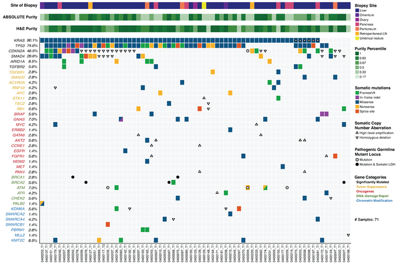
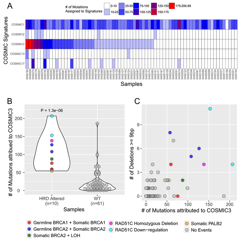
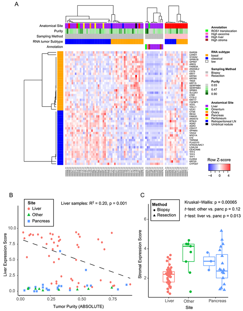
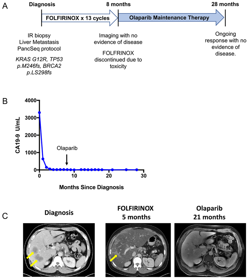
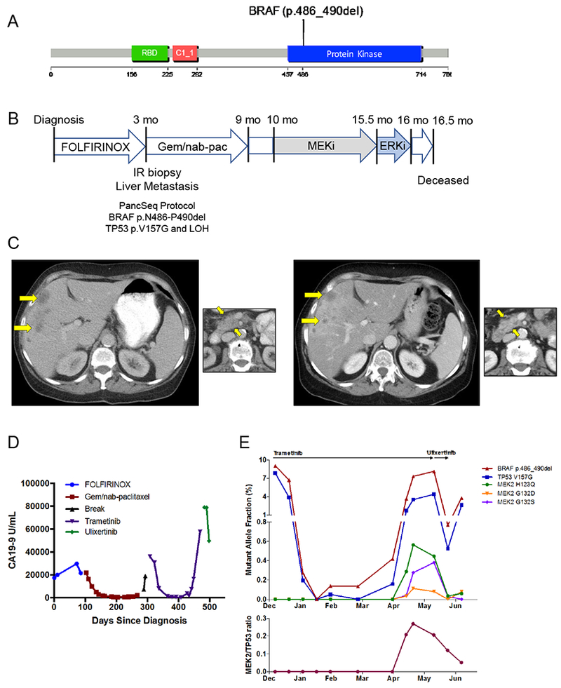
Clinically relevant subtypes exist for pancreatic ductal adenocarcinoma (PDAC), but molecular characterization is not yet standard in clinical care. We implemented a biopsy protocol to perform time-sensitive whole-exome sequencing and RNA sequencing for patients with advanced PDAC. Therapeutically relevant genomic alterations were identified in 48% (34/71) and pathogenic/likely pathogenic germline alterations in 18% (13/71) of patients. Overall, 30% (21/71) of enrolled patients experienced a change in clinical management as a result of genomic data. Twenty-six patients had germline and/or somatic alterations in DNA-damage repair genes, and 5 additional patients had mutational signatures of homologous recombination deficiency but no identified causal genomic alteration. Two patients had oncogenic in-frame BRAF deletions, and we report the first clinical evidence that this alteration confers sensitivity to MAPK pathway inhibition. Moreover, we identified tumor/stroma gene expression signatures with clinical relevance. Collectively, these data demonstrate the feasibility and value of real-time genomic characterization of advanced PDAC.Significance: Molecular analyses of metastatic PDAC tumors are challenging due to the heterogeneous cellular composition of biopsy specimens and rapid progression of the disease. Using an integrated multidisciplinary biopsy program, we demonstrate that real-time genomic characterization of advanced PDAC can identify clinically relevant alterations that inform management of this difficult disease. Cancer Discov; 8(9); 1096-111. ©2018 AACR.See related commentary by Collisson, p. 1062This article is highlighted in the In This Issue feature, p. 1047.


Conflict of interest statement

Conflict of Interest Disclosure: Matthew B. Yurgelun acknowledges research funding from Myriad Genetic Laboratories, Inc. James M. Cleary reports research funding from Merck. Geoffrey I. Shapiro reports research funding from Lilly, Merck, EMD Serono, Sierra Oncology and Pfizer and is a consultant to Pfizer, G1 Therapeutics, Lilly, Roche and Merck/EMD Serono. Charles S. Fuchs is a consultant to CytomX, Sanofi, Eli Lilly, Merck and Entrinsic Health. Levi A. Garraway is an employee of Eli Lilly and Company and has ownership interest in Tango Therapeutic and Foundation Medicine. Bruce E. Johnson reports research funding from Toshiba and Novartis. Nikhil Wagle reports ownership interest in Foundation Medicine. Ryan B. Corcoran reports research funding from AstraZeneca and Sanofi, and is a consultant to Astex, Amgen, Avidity Biosciences, BMS, FOG Pharma, Genentech, LOXO Oncology, Merrimack, N-of-one, Roche, Roivant, Shire, Symphogen, Taiho and WarpDrive Bio. Richard B. Lanham is an employee of and has ownership interest in Guardant Health, Inc. Dean J. Welsch is an employee of BioMed Valley Discoveries. Brian M. Wolpin acknowledges research funding from Celgene. The remaining authors report no potential conflicts of interest.

Figures

Figure 1:
Figure 1:
Landscape of genomic alterations identified by whole exome sequencing in biopsies of advanced pancreatic ductal adenocarcinoma (PDAC) patients. Co-mut plot displaying integrated genomic data for 71 samples displayed as columns, including: somatic mutations, high-level amplifications and homozygous deletions, and germline mutations for selected genes. For each sample, the site of biopsy, the ABSOLUTE neoplastic cellularity (purity) from WES data, and the neoplastic cellularity as assessed by histologic evaluation are shown as tracks at the top. Significantly mutated genes with q value ≤ 0.1 that were identified by exome sequencing are listed at the top (black) vertically in order of decreasing significance. Genes from recurrently altered functional classes are also shown, including tumor suppressor genes (yellow), oncogenes (red), DNA damage repair genes (green) and chromatin modification genes (blue). LOH, loss of heterozygosity. LN, lymph node. Indel, insertion/deletion.
Figure 2:
Figure 2:
Mutational signature analysis of whole exome sequencing data from advanced PDAC patients. A) Projection of signatures representing four main mutational processes identified in de novo signature analysis. All 71 samples in the cohort are listed as columns. Each row represents a signature as defined in the text: COSMIC1 (C>T transitions at CpG dinucleotides, Aging), COSMIC2 and 13 (APOBEC), COSMIC3 (homologous recombination deficiency [HRD] or BRCA deficient), and COSMIC17 (unknown). B) Samples are shown by the number of HRD/COSMIC3 mutations (y-axis) and binned according to whether they have a mutation or gene expression alteration in the known HR genes BRCA1, BRCA2, PALB2 of RAD51C (HRD Altered) (x-axis). Legend at right indicates coloring based on type of alteration. RAD51C downregulation refers to more than 2-fold down-regulation of mRNA-expression levels below the mean value for the entire cohort. “No events” refers to no detected mutation, copy number alteration or mRNA downregulation in the genes indicated. C) Scatter plot of samples displayed by the number of large (≥ 9 base pairs) deletions on the y-axis and the number of HRD/COSMIC3 mutations on the x-axis. Coloring as shown in the legend at right.
Figure 3:
Figure 3:
PDAC gene expression signatures in biopsy cohort. A) Heatmap showing each sample as a column, with rows displaying genes sets defining the Moffitt Basal-like (orange bar) and Classical (blue bar) PDAC gene expression programs (14). Tracks at the top also show anatomic site and ABSOLUTE purity by WES of the sample. To the right of the main biopsy heatmap, samples with low tumor content (middle panel), and samples from resected cases (right most panel) are shown. B) Liver gene expression score (y-axis) is plotted versus the ABSOLUTE purity of each sample (x-axis). The linear regression shown includes only samples from the liver. C) A composite stromal score is displayed for each sample, with samples binned according to the biopsy site. Boxplots represent first, second and third quartiles, and whiskers depict the furthest sample from the median which is within 1.5 times the inter-quartile range.
Figure 4:
Figure 4:
Patient with somatic BRCA2-mutant PDAC demonstrates a radiographic complete response to platinum chemotherapy and subsequent olaparib maintenance therapy. A) 45 year-old man presented with jaundice and abdominal pain and was diagnosed with metastatic PDAC involving the liver and lymph nodes and underwent the depicted treatment course. B) Serum CA19–9 measurements from diagnosis throughout the patient’s treatment course. The arrow indicates transition from FOLFIRINOX chemotherapy to Olaparib (PARP inhibitor) maintenance therapy. C) Computed Tomography (CT) scans are shown at diagnosis demonstrating liver metastases (left panel, yellow arrows) and at the time of cessation of FOLFIRINOX chemotherapy (middle panel) with resolution of liver metastases. Hepatic toxicity of FOLFIRINOX resulted in fatty infiltration of the liver, as noted by severe diffuse attenuation of the liver parenchyma seen in the five month scan. Areas of focal fat sparing in this scan represent treatment effect at the site of liver metastases (middle panel, yellow arrow), denoting a complete response to therapy. The follow-up magnetic resonance imaging (MRI) scan at 21 months after diagnosis, on olaparib maintenance therapy for 13 months, is shown with complete regression of liver metastases (right panel). The patient remains on olaparib therapy without evidence of disease now 28 months after diagnosis.
Figure 5:
Figure 5:
BRAF in-frame deletion confers response to MAPK inhibition. A) An in-frame deletion in BRAF was identified leading to a five amino acid deletion in the kinase domain. B) A 66 year old woman presented with dyspepsia and weight loss and was diagnosed with PDAC and liver metastases and underwent the indicated treatment course. Gem/nab-pac, gemcitabine + nab-paclitaxel. C) CT scans of the liver (large panels) and primary tumor (small panels) at diagnosis (left) and at the time of first restaging scan after 8 weeks of treatment (right) showing partial response to trametinib. D) Serum CA19–9 levels measured throughout the patient’s disease course reflect response and resistance to each therapeutic regimen (color coded by regimen). E) Cell free DNA (cfDNA) measurements for the indicated alleles obtained through droplet digital PCR (ddPCR, BRAF and TP53 alleles) or Guardant360 assay (MEK2 alleles) on plasma collected throughout the patient’s treatment course with trametinib and ulixertinib. Top panel depicts the overall mutant allele fraction of each allele in cfDNA. The bottom panel shows the relative frequency of the MEK2 resistance alleles compared to the overall tumor burden (as measured by the total MEK2/TP53 ratio).

Comment in

References

    1. Rahib L, Smith BD, Aizenberg R, Rosenzweig AB, Fleshman JM, Matrisian LM. Projecting cancer incidence and deaths to 2030: the unexpected burden of thyroid, liver, and pancreas cancers in the United States. Cancer research 2014;74(11):2913–21 doi 10.1158/0008-5472.CAN-14-0155. - DOI - PubMed
    1. Wolfgang CL, Herman JM, Laheru DA, Klein AP, Erdek MA, Fishman EK, et al. Recent progress in pancreatic cancer. CA Cancer J Clin 2013;63(5):318–48 doi 10.3322/caac.21190. - DOI - PMC - PubMed
    1. Ryan DP, Hong TS, Bardeesy N. Pancreatic adenocarcinoma. N Engl J Med 2014;371(11):1039–49 doi 10.1056/NEJMra1404198. - DOI - PubMed
    1. Roberts NJ, Norris AL, Petersen GM, Bondy ML, Brand R, Gallinger S, et al. Whole Genome Sequencing Defines the Genetic Heterogeneity of Familial Pancreatic Cancer. Cancer discovery 2016;6(2):166–75 doi 10.1158/2159-8290.CD-15-0402. - DOI - PMC - PubMed
    1. Sahin IH, Iacobuzio-Donahue CA, O’Reilly EM. Molecular signature of pancreatic adenocarcinoma: an insight from genotype to phenotype and challenges for targeted therapy. Expert Opin Ther Targets 2016;20(3):341–59 doi 10.1517/14728222.2016.1094057. - DOI - PMC - PubMed

Publication types

MeSH terms