

Effect of testosterone treatment on bone remodelling markers and mineral density in obese dieting men in a randomized clinical trial
- PMID: 29904126
- PMCID: PMC6002535
- DOI: 10.1038/s41598-018-27481-3
Effect of testosterone treatment on bone remodelling markers and mineral density in obese dieting men in a randomized clinical trial
Abstract
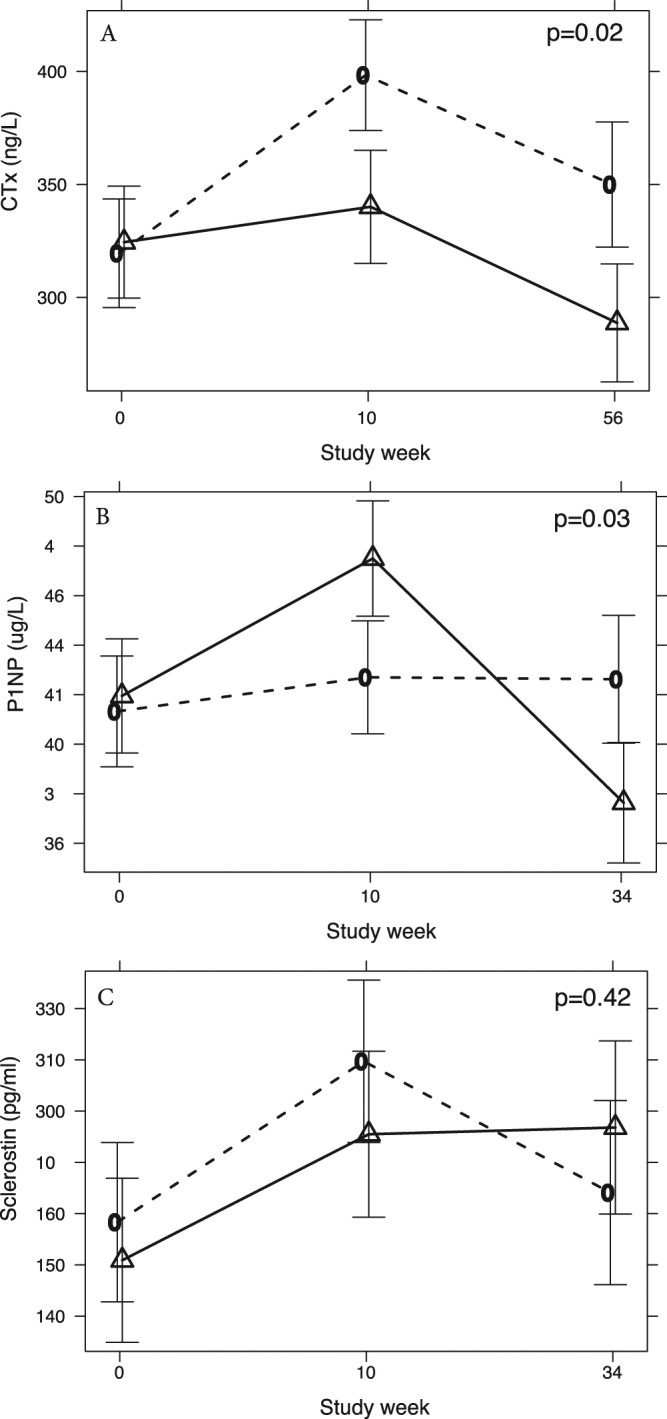
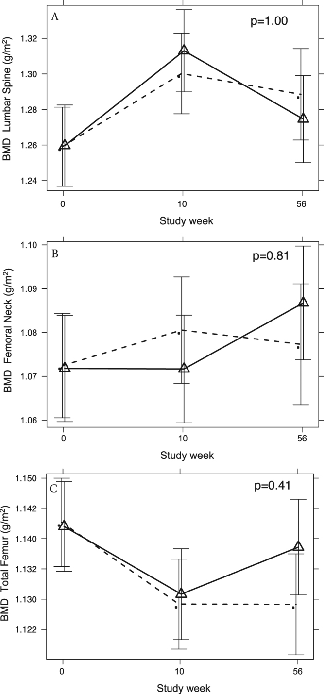
To assess the effect of testosterone treatment on bone remodelling and density in dieting obese men, 100 obese men aged 53 years (interquartile range 47-60) with a total testosterone level <12 nmol/L receiving 10 weeks of a very low energy diet (VLED) followed by 46 weeks of weight maintenance were randomly assigned at baseline to 56 weeks of intramuscular testosterone undecanoate (n = 49, cases) or matching placebo (n = 51, controls). Pre-specified outcomes were between-group differences (mean adjusted difference, MAD) in serum c-telopeptide (CTx), N-terminal propeptide of type 1 procollagen (P1NP) and bone mineral density (BMD). At trial end, CTx was significantly reduced in men receiving testosterone compared to placebo, MAD -66 ng/L (95% CI -113, -18), p = 0.018, and this was apparent already after the 10 week VLED phase, MAD -63 ng/L (95% CI -108, -18), p = 0.018. P1NP was marginally increased after VLED, MAD +4.2 ug/L (95% CI -0.01, +8.4), p = 0.05 but lower at study end, MAD -5.6 ug/L (95% CI -10.1, -1.1), p = 0.03. No significant changes in sclerostin, lumbar spine BMD or femoral BMD were seen. We conclude that in obese men with low testosterone levels undergoing weight loss, bone remodelling markers are modulated in a way that may have favourable effects on bone mass.
Conflict of interest statement
Mathis Grossmann has received research funding from Bayer Pharma, Novartis, Weight Watchers, Lilly and speaker’s honoraria from Besins Healthcare. Mark Ng Tang Fui has received research funding from Bayer Pharma. Rudolf Hoermann, Brendan Nolan, Michelle Clarke and Jeffrey D. Zajac declare that they have no competing interests.
Figures
References
-
- Camacho EM, et al. Age-associated changes in hypothalamic-pituitary-testicular function in middle-aged and older men are modified by weight change and lifestyle factors: longitudinal results from the European Male Ageing Study. Eur. J. Endocrinol. 2013;168:445–455. doi: 10.1530/EJE-12-0890. - DOI - PubMed
-
- Khosla S, et al. Relationship of serum sex steroid levels and bone turnover markers with bone mineral density in men and women: a key role for bioavailable estrogen. J. Clin. Endocrinol. Metab. 1998;83:2266–2274. - PubMed
-
- Barrett-Connor E, et al. Low levels of estradiol are associated with vertebral fractures in older men, but not women: the Rancho Bernardo Study. J. Clin. Endocrinol. Metab. 2000;85:219–223. - PubMed
Publication types
MeSH terms
Substances
LinkOut - more resources
Full Text Sources
Other Literature Sources
Medical
Research Materials
Miscellaneous
