. 2018 Sep;23(9):1851-1867.
doi: 10.1038/s41380-018-0100-y. Epub 2018 Jun 14.

Diazepam-induced loss of inhibitory synapses mediated by PLCδ/ Ca2+/calcineurin signalling downstream of GABAA receptors

Affiliations

Diazepam-induced loss of inhibitory synapses mediated by PLCδ/ Ca2+/calcineurin signalling downstream of GABAA receptors

Martin W Nicholson et al. Mol Psychiatry. 2018 Sep.

Abstract

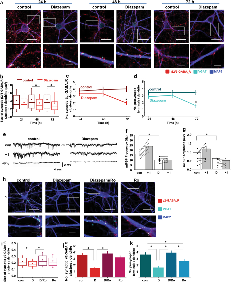
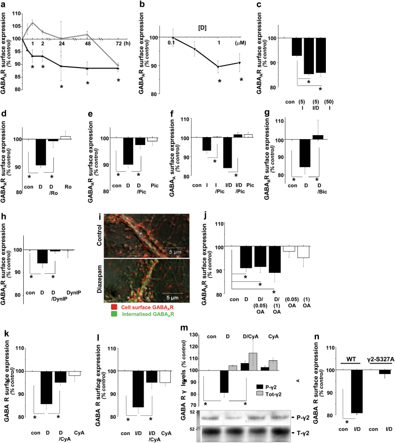
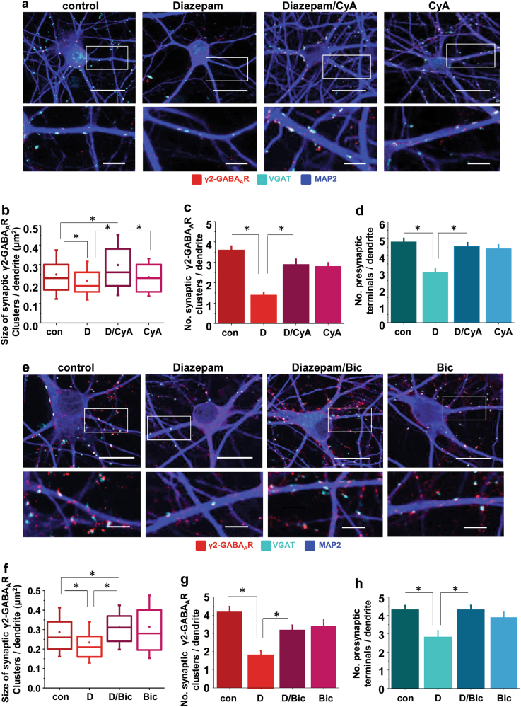
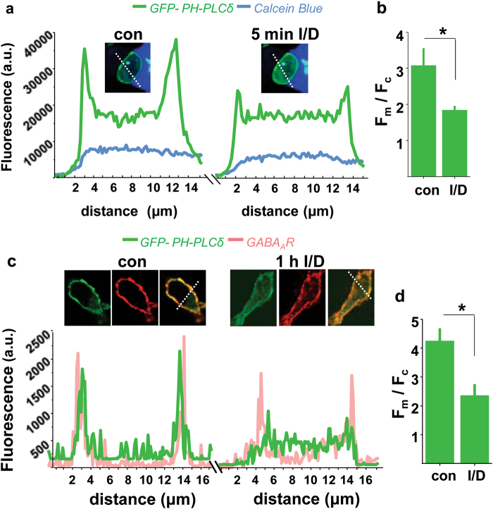
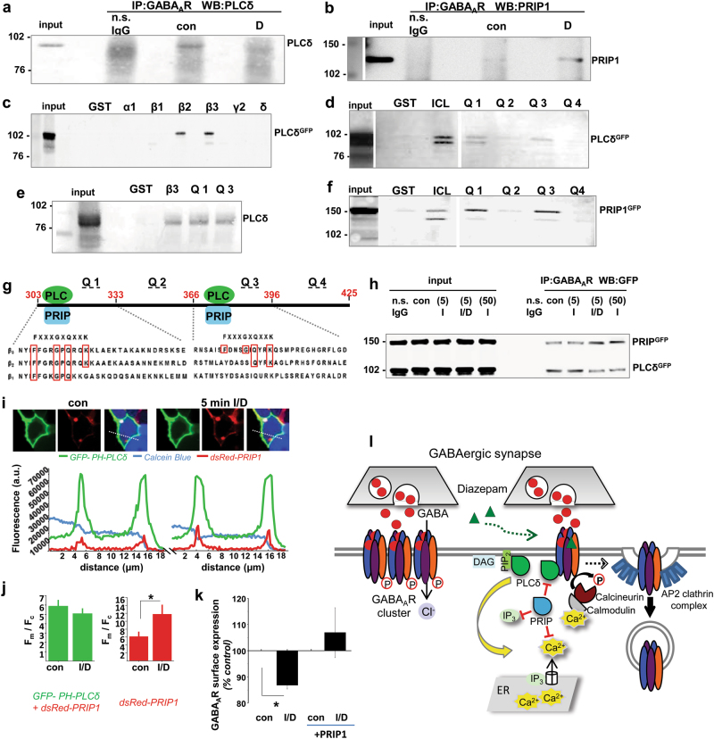
Benzodiazepines facilitate the inhibitory actions of GABA by binding to γ-aminobutyric acid type A receptors (GABAARs), GABA-gated chloride/bicarbonate channels, which are the key mediators of transmission at inhibitory synapses in the brain. This activity underpins potent anxiolytic, anticonvulsant and hypnotic effects of benzodiazepines in patients. However, extended benzodiazepine treatments lead to development of tolerance, a process which, despite its important therapeutic implications, remains poorly characterised. Here we report that prolonged exposure to diazepam, the most widely used benzodiazepine in clinic, leads to a gradual disruption of neuronal inhibitory GABAergic synapses. The loss of synapses and the preceding, time- and dose-dependent decrease in surface levels of GABAARs, mediated by dynamin-dependent internalisation, were blocked by Ro 15-1788, a competitive benzodiazepine antagonist, and bicuculline, a competitive GABA antagonist, indicating that prolonged enhancement of GABAAR activity by diazepam is integral to the underlying molecular mechanism. Characterisation of this mechanism has revealed a metabotropic-type signalling downstream of GABAARs, involving mobilisation of Ca2+ from the intracellular stores and activation of the Ca2+/calmodulin-dependent phosphatase calcineurin, which, in turn, dephosphorylates GABAARs and promotes their endocytosis, leading to disassembly of inhibitory synapses. Furthermore, functional coupling between GABAARs and Ca2+ stores was sensitive to phospholipase C (PLC) inhibition by U73122, and regulated by PLCδ, a PLC isoform found in direct association with GABAARs. Thus, a PLCδ/Ca2+/calcineurin signalling cascade converts the initial enhancement of GABAARs by benzodiazepines to a long-term downregulation of GABAergic synapses, this potentially underpinning the development of pharmacological and behavioural tolerance to these widely prescribed drugs.


Conflict of interest statement

The authors declare that they have no conflict of interest.

Figures

Fig. 1
Fig. 1
Diazepam causes a time-dependent breakdown of GABAergic inhibitory synapses upon direct binding to GABAARs. a Immunolabeling of postsynaptic GABAAR β2/3-containing clusters (red) and VGAT-positive presynaptic GABAergic terminals (cyan) along MAP2-positive primary dendrites (20 µm; blue) of cortical neurons in the absence or presence of diazepam (D; 1 μM), and the corresponding graphs showing a decrease over time in b size (median/line-IQRs; mean/dot ± s.d. whiskers; Mann–Whitney test, *p<0.05) and c number (mean ± s.e.m.; ANOVA/Bonferonni post-hoc test; *p<0.05) of synaptic β2/3 clusters, and d number of GABAergic terminals (mean ± s.e.m.; ANOVA/Bonferonni post-hoc test; *p<0.05) contacting n=59, n=70, n=53 control (DMSO)-treated primary dendrites and n = 70, n = 67, n = 44 diazepam-treated primary dendrites for 24 h, 48 h, and 72 h, respectively. Total of n = 17, n = 18 and n = 15 control and n = 17, n = 17 and n = 17 diazepam-treated neurons, respectively, collected from two independent experiments, were analysed in each group. e Representative traces of mIPSPs recorded in cortical neurons after 72 h treatment with control (DMSO) or diazepam (D; 1 μM), before and after application of isoguvacine ( + I, 50 μM), followed by picrotoxin ( + Pic, 50 μM; scale refers to all conditions), and corresponding bar graphs (mean ± s.d.; Student’s t-test: *p < 0.05) showing a diazepam-dependent decrease in f frequency and g amplitude of mIPSPs and the effects of isoguvacine ( + I, 50 μM; grey bars) from n = 7 control & n = 7 diazepam-treated cells collected from n = 3 independent experiments. h Immunolabelling of γ2-containing clusters (red) and VGAT-GABArgic terminals (cyan) along MAP2-positive dendrites (20 µm; blue) following 72 h treatment with control (DMSO), or diazepam (D; 1 μM), in the absence or presence of Ro 15-1788 (Ro; 25 μM), and the corresponding graphs showing a decrease in i size (median/line-IQRs; mean/dot ± s.d. whiskers; Mann–Whitney test, *p < 0.05), and j number of synaptic γ2 clusters (mean ± s.e.m.; ANOVA/Bonferonni post-hoc test; *p < 0.05), and k decrease in the number of GABAergic terminals (mean ± s.e.m.; ANOVA/Bonferonni post-hoc test; *p< 0.05) contacting n = 49 control-treated & n = 45, n = 53, n = 48 diazepam-, diazepam/Ro- or Ro-treated dendrites for 72 h, respectively. The total of n = 15, n = 14, n= 13, n = 16 neurons, respectively, collected from two independent experiments, were analysed in each group. Scale bars = 20 μm (a, h-upper row) and = 5 μm (a, h-lower row)
Fig. 2
Fig. 2
Time and dose-dependent internalisation of GABAARs in response to diazepam requires calcineurin activity. a Diazepam (D; 1 μM)-dependent decrease in surface (black) and total (grey) levels of GABAARs over time (n=5), and b in surface levels only in the presence of increasing doses for 2 h (n=4) in cortical neurones. c Decrease in surface GABAARs in response to low doses of isoguvacine (I; 5 μM) is potentiated by diazepam (D; 1 μM) in α122myc-HEK293 cell line (n=6). dg Diazepam (D; 1 μM)-dependent decrease in surface GABAARs is inhibited by Ro 15-1788 (Ro; 25 μM; d; n=7), picrotoxin (Pic; 50 μM; e; n=5), or bicuculline (Bic; 50 μM; g; n=4) in cortical neurones, and f picrotoxin (Pic; 50 μM; n=6) in α122myc-HEK293 cell line. h Diazepam (D; 1 μM)-dependent decrease in surface GABAARs is inhibited by dynamin-inhibitory peptide (DynIP; 25 μM; n=5) in cortical neurones following 2 h treatments. i Immunolabelling of internalised (green) and surface (red) GABAARs following 2 h treatments with diazepam (D; 1 μM; n=2; scale bar = 5 µm). j Diazepam (D; 1 μM)-dependent reduction of surface GABAARs is unaffected by inhibition of PP2A or PP1 with low (0.05 μM) or high (1 μM) dose of okadaic acid, respectively (OA; n=6), but k it is prevented by inhibition of calcineurin by cyclosporine A (CyA; 1 μM; n=6). l Diazepam (D; 1 μM) /isoguvacine (I; 5 μM)-dependent reduction in surface GABAARs in α122myc-HEK293 cell line is prevented by inhibition of calcineurin with cyclosporine A (CyA; 1 μM; n=3). m Diazepam treatments (D; 1 μM; 2 h) cause GABAAR γ2 subunit dephosphorylation at Ser327 in cortical neurones and this is prevented by cyclosporine A (CyA; 1 μM; n=2). Immunoblotting was done using an anti-PSer327-γ2 or anti-γ2 primary antibody, followed by alkaline phosphatase-conjugated secondary antibody and a colour reaction. Quantification was done using ImageJ. n Diazepam (D; 1 μM)/isoguvacine (I; 5 μM)-dependent reduction in surface GABAARs in α122myc-HEK293 cells is abolished by S327A mutation in the γ2 subunit. Changes in surface GABAARs were measured by cell surface ELISA using β2/3-specific antibody in cortical neurones or myc-antibody in α122myc-HEK293 cells and presented in graphs as mean ± s.e.m., with n = number of independent experiments. Statistical analysis was done using ANOVA with Bonferonni post-hoc test; *p < 0.05
Fig. 3
Fig. 3
Diazepam-dependent loss of GABAergic synapses is prevented by inhibition of calcineurin or GABAAR activity. a Immunolabelling of γ2-containing clusters (red) and VGAT-positive presynaptic GABArgic terminals (cyan) along MAP2-positive dendrites (20 µm; blue) following 72 h treatment with control (DMSO) or diazepam (D; 1 μM), in the absence or presence of cyclosporine A (CyA; 1 μM), and corresponding graphs showing b size (median/line-IQRs; mean/dot ± s.d. whiskers; Mann–Whitney test, *p < 0.05), and c number of synaptic γ2 clusters (mean ± s.e.m.; ANOVA/Bonferonni post-hoc test; *p < 0.05), from n = 71 control dendrites & n = 85, n = 64, n = 73 dendrites of diazepam-, diazepam/cyclosporine A and cyclosporine A-treated cells, respectively, of a total of n=19, n=19, n=18, n=19 neurons in each group collected from two independent experiments. d Decrease in number of GABAergic terminals (mean ± s.e.m.; ANOVA/Bonferonni post-hoc test; *p < 0.05) contacting n=71 control-treated & n=85, n=64, n=73 diazepam-, diazepam/cyclosporine A- or cyclosporine A-treated dendrites for 72 h, respectively, from a total of n=19, n=19, n=18, n= 19 neurons in each group, collected from two independent experiments. e Immunolabelling of γ2-containing clusters (red) and VGAT-positive presynaptic GABArgic terminals (cyan) along MAP2-positive dendrites (20 µm; blue) following 72 h treatment with control (DMSO) or diazepam (D; 1 μM), in the absence or presence of bicuculline (Bic; 50 μM), and corresponding graphs showing f size (median/line-IQRs; mean/dot ± s.d. whiskers; Mann–Whitney test, *p < 0.05), and g number of synaptic γ2 clusters (mean ± s.e.m.; ANOVA/Bonferonni post-hoc test; *p<0.05), from n= 60 control dendrites & n= 63, n= 61, n = 65 dendrites of diazepam-, diazepam/bicuculline and bicuculline-treated cells, respectively, of a total of n = 16, n = 19, n = 18, n = 20 neurons in each group collected from two independent experiments. h Decrease in number of GABAergic terminals (mean ± s.e.m.; ANOVA/Bonferonni post-hoc test; *p<0.05), contacting n = 60 control-treated & n = 63, n = 61, n= 65 diazepam-, diazepam/bicuculine- or bicuculline-treated dendrites for 72 h, respectively, from a total of n = 16, n = 19, n = 18, n = 20 neurons in each group, collected from two independent experiments. Scale bars = 20 μm (a, e-upper row) and = 5 μm (a, h-lower row)
Fig. 4
Fig. 4
Diazepam triggers release of Ca2+ from the intracellular stores which is required for internalisation of GABAARs and prevented by Ro 15-1788, bicuculline, thapsigargin and U-73122. Time lapse imaging of intracellular Ca2+ in Fluo-4-labelled cortical neurones treated with diazepam (D; 1 μM) alone (a, inset: representative images before and after diazepam addition; scale bars = 20 μm), or in the presence of Ro15-1788 (Ro, 25 μM; b) or bicuculine (Bic, 50 μM; c), shown as a fluorescence ratio Ft/F0, and d quantified at the peak of response to diazepam in dendrites and somas (d; mean ± s.e.m.; ANOVA/Bonferonni post-hoc test; *p<0.05; n=5 neurones in each group from 2 independent experiments). e Diazepam (D; 1 μM)-dependent increase in intracellular Ca2+ (insert: representative images before and after diazepam addition; scale bars = 20 μm) is inhibited by thapsigargin (T; 2 μM; f) and U-73122 (U; 10 μM; g). h Ft/F0 was quantified at the peak of response to diazepam in dendrites and somas of labelled cortical neurons (mean ± s.e.m.; ANOVA/Bonferonni post-hoc test; *p< 0.05; n = 4 neurones in each group from 2 independent experiments). i–k Diazepam (D; 1 μM)-dependent internalisation of GABAARs in neurones (left), and Diazepam (D; 1 μM)/Isoguvacine (I; 5 μM)-dependent internalisation of GABAARs in α122myc-HEK293 cells (right) are prevented by thapsigargin (T; 2 μM; i) and U-73122 (U; 10 μM; j), but insensitive to EGTA (E; 1 mM; k). Changes in surface GABAARs were measured by cell surface ELISA using β2/3-specific antibody in neurones or myc-antibody in α122myc-HEK293 cells and presented in graphs as mean ± s.e.m. Statistical analysis was done using ANOVA with Bonferonni post-hoc test; *p<0.05 (n=7 thapsigargin, n = 5 U-73122, n = 9 EGTA independent experiments)
Fig. 5
Fig. 5
Diazepam/Isoguvacine-dependent translocation of GFP-PHPLCδ1 from the cell membrane to the cytoplasm in α122-HEK293 cells. a Live imaging of a Calcein blue-labelled cell (inset) showing changes in GFP-PHPLCδ1 fluorescence intensity profile (green) prior to (left) and 5 min after the addition of Diazepam (D; 1 μM)/Isoguvacine (I; 5 μM) (right). b Quantification of fluorescence F(membrane)/F(cytoplasm) ratio of GFP-PHPLCδ1 (green; mean ± s.e.m.; Student t-test; *p < 0.05; n= 10 cells from 2 independent experiments). c Imaging of surface GABAAR-β2-subunit in fixed HEK293 cells (red) expressing GFP-PHPLCδ1 (green) in control (DMSO, left) and Diazepam (D; 1 μM)/Isoguvacine (I; 5 μM) treated samples for 1 h (right), showing superimposed fluorescence intensity profiles across the selected cells. d Quantification of fluorescence F(membrane)/F(cytoplasm) ratio of GFP-PHPLCδ1 (green; mean ± s.e.m.; Student t-test; *p < 0.05; n = 10 cells from 2 independent experiments)
Fig. 6
Fig. 6
Diazepam triggers dissociation of PLCδ from GABAARs in situ leading to activation of PLCδ/Ca2+/calcineurin signalling pathway, which is negatively regulated by PRIP1. a Immunoprecipitates of GABAARs from control and diazepam (D; 1 μM)-treated cortical neurones were probed with PLCδ- (n= 3) or b PRIP1-  (n = 4) specific antibody. ce PLCδ-GFP binds directly to the intracellular loop of the GABAAR β2 and β3 subunits Q1 (303-366 aa) and Q3 (366-396 aa) regions in GST pull-down assays (n = 3). f PRIP1-GFP binds directly to the β3 subunit Q1 and Q3 loop regions in the GST pull-down assays (n = 3). g Predicted PLCδ- and PRIP1- binding sites in the Q1 and Q3 regions of the β2 and β3 subunits. h Immunoprecipitates of GABAARs from control (DMSO) or Isoguvacine (I; 5 μM)-, Diazepam (D; 1 μM)/Isoguvacine (I; 5 μM)- or Isoguvacine (I; 50 μM)-treated α1β2γ2-GABAAR HEK293 cells expressing both GFP-PLCδ and GFP-PRIP1 were probed with the GFP-specific antibody (n = 2). (i) Overexpression of PRIP1 inhibits partial translocation of GFP-PHPLCδ1 in response to Diazepam (D; 1 μM)/Isoguvacine (I; 5 μM) in α1β2γ2-HEK293 cells. Live imaging of a Calcein blue-labelled cell (blue) expressing GFP-PHPLCδ1 (green) and dsRed-PRIP1 (red; top panels) and superimposed fluorescence intensity profiles prior to (left) and 5 min after the addition of Diazepam (D; 1 μM)/Isoguvacine (I; 5 μM) (right). j Quantification of fluorescence F(membrane)/F(cytoplasm) ratio of GFP-PHPLCδ1 (green; left) and dsRed-PRIP1 (red; right), both shown as mean ± s.e.m. (Student t-test; *p < 0.05; n = 10 cells from 2 independent experiments). k Overexpression of PRIP1 inhibits Diazepam (D; 1 μM)/Isoguvacine (I; 5 μM)-dependent internalisation of GABAARs. Changes in surface GABAARs were measured by cell surface ELISA with anti-myc-specific antibody labelling the γ2 subunit, and presented as mean ± s.e.m. (n = 4). Statistical analysis was done using ANOVA with Bonferonni post-hoc test; *p<0.05; n = number of independent experiments. l Schematic diagram of the GABAAR/PLCδ/Ca2+/calcineurin feed-back mechanism underlying diazepam-dependent downregulation of GABAARs. According to this model, sustained activation of synaptic GABAARs by diazepam triggers a metabotropic, PLCδ/Ca2+/calcineurin signalling pathway which leads to receptor dephosphorylation by calcineurin, initiation of dynamin-dependent endocytosis resulting in a decrease in the size and number of postsynaptic GABAAR clusters, and disassembly of inhibitory synapses. This mechanism is ‘switched off’ when PRIP1, PLCδ-related but catalytically inactive protein, outcompetes the PLCδ in binding to GABAARs, thereby preventing the activation of PLCδ and downstream Ca2+/calcineurin-dependent internalisation of these receptors

Similar articles

Cited by

References

    1. Rudolph U, Knoflach F. Beyond classical benzodiazepines: novel therapeutic potential of GABAA receptor subtypes. Nat Rev Drug Discov. 2011;10:685–97. doi: 10.1038/nrd3502. - DOI - PMC - PubMed
    1. Spiers N, Qassem T, Bebbington P, McManus S, King M, Jenkins R, et al. Prevalence and treatment of common mental disorders in the English national population, 1993–2007. Br J Psychiatry. 2016;209:150–6. doi: 10.1192/bjp.bp.115.174979. - DOI - PubMed
    1. Lader M. Benzodiazepine harm: how can it be reduced? Br J Clin Pharmacol. 2014;77:295–301. doi: 10.1111/j.1365-2125.2012.04418.x. - DOI - PMC - PubMed
    1. Lugoboni F, Quaglio G. Exploring the dark side of the moon: the treatment of benzodiazepine tolerance. Br J Clin Pharmacol. 2014;77:239–41. doi: 10.1111/bcp.12148. - DOI - PMC - PubMed
    1. Downing SS, Lee YT, Farb DH, Gibbs TT. Benzodiazepine modulation of partial agonist efficacy and spontaneously active GABA(A) receptors supports an allosteric model of modulation. Br J Pharmacol. 2005;145:894–906. doi: 10.1038/sj.bjp.0706251. - DOI - PMC - PubMed

Publication types

MeSH terms