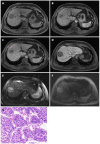
Noninvasive imaging of hepatocellular carcinoma: From diagnosis to prognosis
- PMID: 29904242
- PMCID: PMC6000290
- DOI: 10.3748/wjg.v24.i22.2348
Noninvasive imaging of hepatocellular carcinoma: From diagnosis to prognosis
Abstract
Hepatocellular carcinoma (HCC) is the most common primary liver cancer and a major public health problem worldwide. Hepatocarcinogenesis is a complex multistep process at molecular, cellular, and histologic levels with key alterations that can be revealed by noninvasive imaging modalities. Therefore, imaging techniques play pivotal roles in the detection, characterization, staging, surveillance, and prognosis evaluation of HCC. Currently, ultrasound is the first-line imaging modality for screening and surveillance purposes. While based on conclusive enhancement patterns comprising arterial phase hyperenhancement and portal venous and/or delayed phase wash-out, contrast enhanced dynamic computed tomography and magnetic resonance imaging (MRI) are the diagnostic tools for HCC without requirements for histopathologic confirmation. Functional MRI techniques, including diffusion-weighted imaging, MRI with hepatobiliary contrast agents, perfusion imaging, and magnetic resonance elastography, show promise in providing further important information regarding tumor biological behaviors. In addition, evaluation of tumor imaging characteristics, including nodule size, margin, number, vascular invasion, and growth patterns, allows preoperative prediction of tumor microvascular invasion and patient prognosis. Therefore, the aim of this article is to review the current state-of-the-art and recent advances in the comprehensive noninvasive imaging evaluation of HCC. We also provide the basic key concepts of HCC development and an overview of the current practice guidelines.
Keywords: Computed tomography; Diagnosis; Guidelines; Hepatocellular carcinoma; Magnetic resonance imaging; Prognosis; Staging; Surveillance; Ultrasound.
Conflict of interest statement
Conflict-of-interest statement: All authors declared no potential conflict of interests related to this publication.
Figures


Similar articles
-
Recent advances in non-invasive magnetic resonance imaging assessment of hepatocellular carcinoma.World J Gastroenterol. 2018 Jun 21;24(23):2413-2426. doi: 10.3748/wjg.v24.i23.2413. World J Gastroenterol. 2018. PMID: 29930464 Free PMC article. Review.
-
Emerging Role of Hepatobiliary Magnetic Resonance Contrast Media and Contrast-Enhanced Ultrasound for Noninvasive Diagnosis of Hepatocellular Carcinoma: Emphasis on Recent Updates in Major Guidelines.Korean J Radiol. 2019 Jun;20(6):863-879. doi: 10.3348/kjr.2018.0450. Korean J Radiol. 2019. PMID: 31132813 Free PMC article. Review.
-
Image fusion with volume navigation of contrast enhanced ultrasound (CEUS) with computed tomography (CT) or magnetic resonance imaging (MRI) for post-interventional follow-up after transcatheter arterial chemoembolization (TACE) of hepatocellular carcinomas (HCC): Preliminary results.Clin Hemorheol Microcirc. 2010;46(2-3):101-15. doi: 10.3233/CH-2010-1337. Clin Hemorheol Microcirc. 2010. PMID: 21135486
-
Diagnosis and staging of hepatocellular carcinoma (HCC): current guidelines.Eur J Radiol. 2018 Apr;101:72-81. doi: 10.1016/j.ejrad.2018.01.025. Epub 2018 Jan 31. Eur J Radiol. 2018. PMID: 29571804 Review.
-
Evaluation of hemodynamic imaging findings of hypervascular hepatocellular carcinoma: comparison between dynamic contrast-enhanced magnetic resonance imaging using radial volumetric imaging breath-hold examination with k-space-weighted image contrast reconstruction and dynamic computed tomography during hepatic arteriography.Jpn J Radiol. 2018 Apr;36(4):295-302. doi: 10.1007/s11604-018-0720-9. Epub 2018 Jan 11. Jpn J Radiol. 2018. PMID: 29327116
Cited by
-
Prognostic value of AFP-L3 and Des-γ-carboxy prothrombin in advanced primary liver cancer treated with Sorafenib and transarterial chemoembolization.Am J Transl Res. 2024 Sep 15;16(9):5004-5010. doi: 10.62347/PMYP4404. eCollection 2024. Am J Transl Res. 2024. PMID: 39398579 Free PMC article.
-
A Rare Presentation of Hepatocellular Carcinoma Infiltrating the Gallbladder.Cureus. 2019 Jul 15;11(7):e5140. doi: 10.7759/cureus.5140. Cureus. 2019. PMID: 31523569 Free PMC article.
-
A Comprehensive Overview of the Efficacy and Safety of Gadopiclenol: A New Contrast Agent for MRI of the CNS and Body.Invest Radiol. 2024 Feb 1;59(2):124-130. doi: 10.1097/RLI.0000000000001025. Epub 2023 Oct 9. Invest Radiol. 2024. PMID: 37812485 Free PMC article. Review.
-
An Optimized Integrin α6-Targeted Magnetic Resonance Probe for Molecular Imaging of Hepatocellular Carcinoma in Mice.J Hepatocell Carcinoma. 2021 Jun 24;8:645-656. doi: 10.2147/JHC.S312921. eCollection 2021. J Hepatocell Carcinoma. 2021. PMID: 34235103 Free PMC article.
-
Aptamer-functionalized nanomaterials (AFNs) for therapeutic management of hepatocellular carcinoma.J Nanobiotechnology. 2024 May 12;22(1):243. doi: 10.1186/s12951-024-02486-5. J Nanobiotechnology. 2024. PMID: 38735927 Free PMC article. Review.
References
-
- International Agency for Research on Cancer. GLOBOCAN 2012: Esti-mated cancer incidence, mortaloty and prevalence worldwide in 2012. Accessed to 2018-02-28. Available from: http://globocan.iarc.fr/old/FactSheets/cancers/liver-new.asp.
-
- Forner A, Llovet JM, Bruix J. Hepatocellular carcinoma. Lancet. 2012;379:1245–1255. - PubMed
-
- Dimitroulis D, Damaskos C, Valsami S, Davakis S, Garmpis N, Spartalis E, Athanasiou A, Moris D, Sakellariou S, Kykalos S, et al. From diagnosis to treatment of hepatocellular carcinoma: An epidemic problem for both developed and developing world. World J Gastroenterol. 2017;23:5282–5294. - PMC - PubMed
-
- El-Serag HB. Hepatocellular carcinoma. N Engl J Med. 2011;365:1118–1127. - PubMed
Publication types
MeSH terms
Substances
LinkOut - more resources
Full Text Sources
Other Literature Sources
Medical

