. 2018 May 31:11:182.
doi: 10.3389/fnmol.2018.00182. eCollection 2018.

Medium-Chain Fatty Acids, Beta-Hydroxybutyric Acid and Genetic Modulation of the Carnitine Shuttle Are Protective in a Drosophila Model of ALS Based on TDP-43

Affiliations

Medium-Chain Fatty Acids, Beta-Hydroxybutyric Acid and Genetic Modulation of the Carnitine Shuttle Are Protective in a Drosophila Model of ALS Based on TDP-43

Ernesto Manzo et al. Front Mol Neurosci. .

Abstract

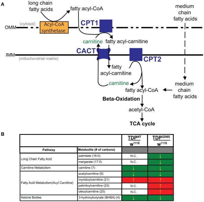
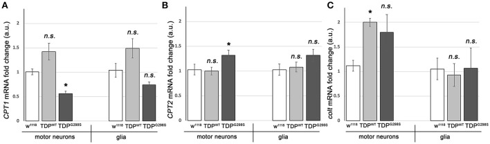
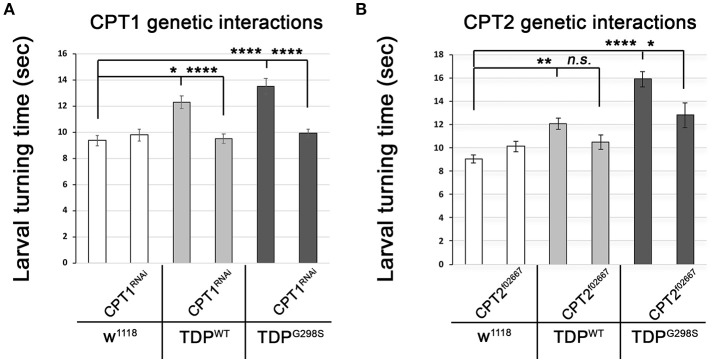
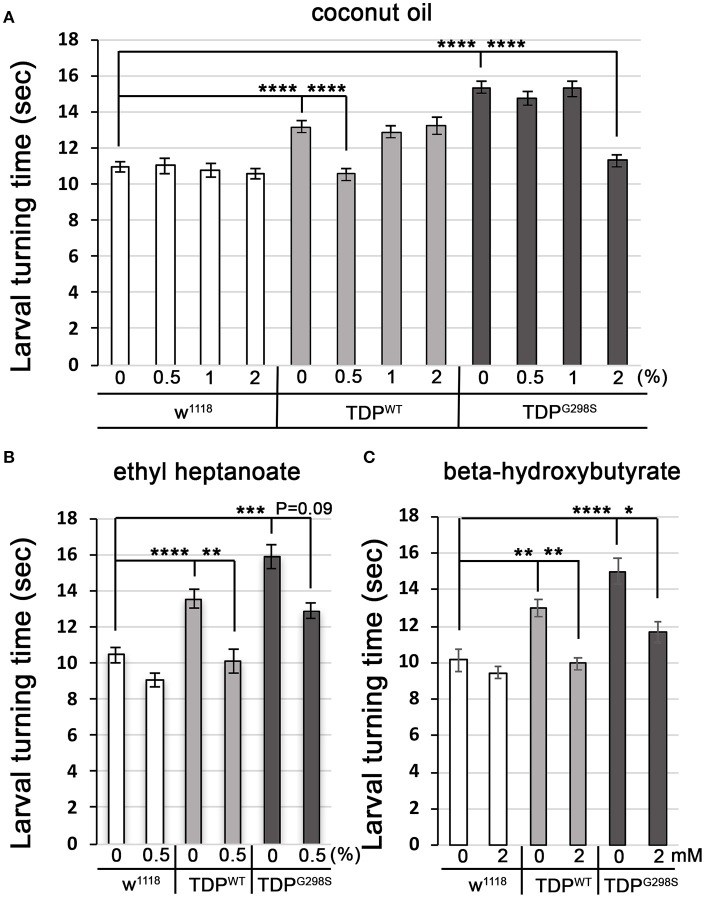
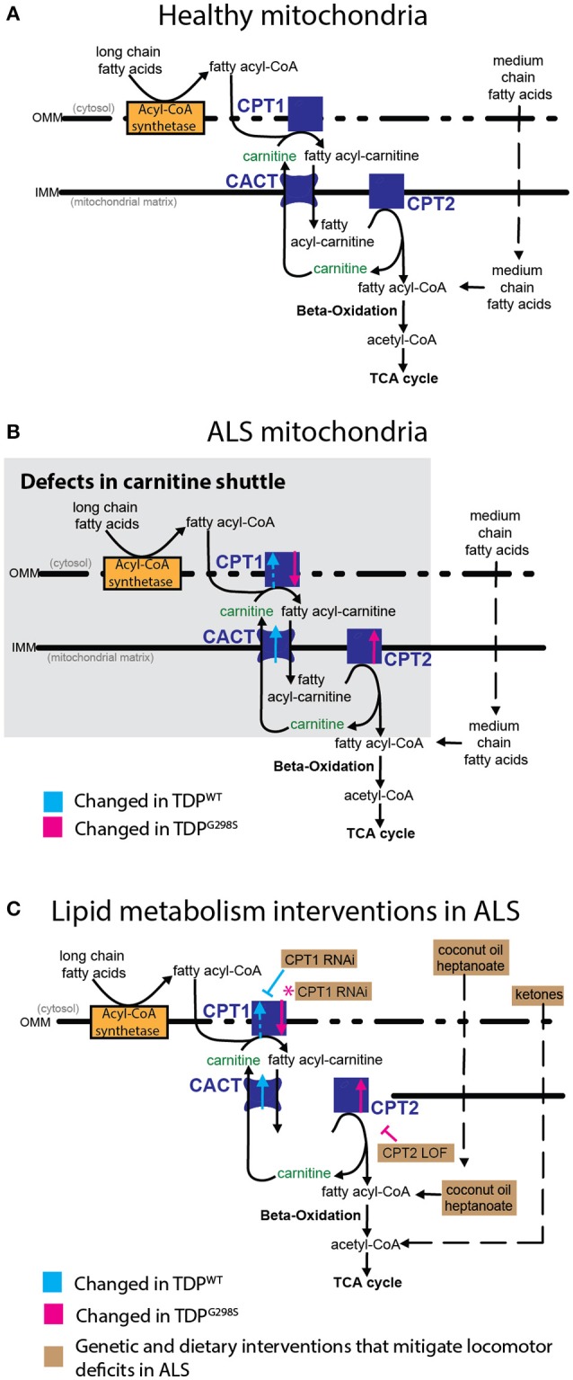
ALS patients exhibit dyslipidemia, hypermetabolism and weight loss; in addition, cellular energetics deficits have been detected prior to denervation. Although evidence that metabolism is altered in ALS is compelling, the mechanisms underlying metabolic dysregulation and the contribution of altered metabolic pathways to disease remain poorly understood. Here we use a Drosophila model of ALS based on TDP-43 that recapitulates hallmark features of the disease including locomotor dysfunction and reduced lifespan. We performed a global, unbiased metabolomic profiling of larvae expressing TDP-43 (wild-type, TDPWT or disease-associated mutant, TDPG298S) and identified several lipid metabolism associated alterations. Among these, we found a significant increase in carnitine conjugated long-chain fatty acids and a significant decrease in carnitine, acetyl-carnitine and beta-hydroxybutyrate, a ketone precursor. Taken together these data suggest a deficit in the function of the carnitine shuttle and reduced lipid beta oxidation. To test this possibility we used a combined genetic and dietary approach in Drosophila. Our findings indicate that components of the carnitine shuttle are misexpressed in the context of TDP-43 proteinopathy and that genetic modulation of CPT1 or CPT2 expression, two core components of the carnitine shuttle, mitigates TDP-43 dependent locomotor dysfunction, in a variant dependent manner. In addition, feeding medium-chain fatty acids or beta-hydroxybutyrate improves locomotor function, consistent with the notion that bypassing the carnitine shuttle deficit is neuroprotective. Taken together, our findings highlight the potential contribution of the carnitine shuttle and lipid beta oxidation in ALS and suggest strategies for therapeutic intervention based on restoring lipid metabolism in motor neurons.

Keywords: TDP-43; amyotrophic lateral sclerosis; beta lipid oxidation; carnitine shuttle; lipid metabolism; metabolomics.


Figures

Figure 1
Figure 1
Metabolomic profiling of ALS larvae uncovers deficits in carnitine, carnitine conjugation and lipid beta oxidation. (A) Carnitine shuttle components and fatty acid import into mitochondria. CPT1, 2, carnitine acyltransferases; CACT, carnitine acylcarnitine transferase; IMM, Inner mitochondrial membrane; OMM, Outer mitochondrial membrane. (B) Summary of significantly altered metabolites in TDPWT and TDPG298S compared to w1118 controls. Green indicates significant downregulation and red indicates significant upregulation (Pvalue < 0.05).
Figure 2
Figure 2
TDP-43 causes alterations in the expression of carnitine shuttle components. (A–C) Transcriptional profiling using q RT PCR of CPT1 mRNA (A), CPT2 mRNA (B) and colt mRNA (C) indicates TDP-43 dependent changes in motor neurons and glia. TDPWT or TDPG298S were expressed using D42 GAL4 (motor neurons) or repo GAL4 (glia). Kruskal-Wallis multiple comparisons test was used to calculate significance. n.s., not significant; *Pvalue < 0.05.
Figure 3
Figure 3
Genetic interaction experiments show that CPT1 and CPT2 mitigate locomotor dysfunction caused by TDPWT and TDPG298S, in a variant dependent manner. (A) CPT1 knock-down by RNAi (CPT1RNAi, using y1v1; P{y[+t7.7] v[+t1.8] = TRiP.HMS00040}attP2/TM3, Sb1) mitigates TDPWT and TDPG298S larval turning times. (B) CPT2 loss of function (CPT2f02667, using w1118; PBac{w[+mC]=WH}CPT2f02667/TM6B, Tb1) mitigates TDPG298S larval turning times. TDPWT or TDPG298S were expressed in motor neurons using D42 GAL4. Genotypes as indicated. Kruskal-Wallis multiple comparisons test was used to calculate significance. *Pvalue < 0.05, **Pvalue < 0.01, ****Pvalue < 0.0001.
Figure 4
Figure 4
Medium-chain fatty acids and ketones mitigate TDP-43 dependent locomotor dysfunction in a Drosophila model of ALS. (A,B) Larval turning times, used to measure locomotor deficits caused by TDPWT or TDPG298S, are rescued by feeding different medium-chain fatty acids [coconut oil (A) or ethyl heptanoate (B)]. (C) Ketone (beta-hydroxybutyrate) feeding reduces larval turning times in ALS larvae. TDPWT or TDPG298S were expressed in motor neurons using D42 GAL4. Genotypes and treatments as indicated. Kruskal-Wallis multiple comparisons test was used to calculate significance. *Pvalue < 0.05, **Pvalue < 0.01, ***Pvalue < 0.001, ****Pvalue < 0.0001.
Figure 5
Figure 5
Model of mitochondrial dysfunction in ALS and proposed interventions. Expression of TDPWT or TDPG298S in motor neurons causes alterations in the expression of carnitine shuttle components (ALS mitochondria, B) compared to controls (Healthy mitochondria, A). Blue dashed line for CPT1 indicates an upward trend in transcript levels (Pvalue = 0.08). These defects can be countered genetically, by reducing CPT1 in the context of TDPWT (inhibitory blue symbol), or reducing CPT2 in the context of TDPG298S (inhibitory magenta symbol). Magenta asterisk indicates a potential compensatory mechanism whereby CPT1 RNAi knock-down mitigates TDPG298S locomotor phenotypes despite CPT1 mRNA being reduced. Alternatively, dietary supplementation with medium-chain fatty acids or beta-hydroxybutyrate bypasses carnitine shuttle dysfunction leading to significant improvement in locomotor function (C, Lipid metabolism interventions in ALS).

References

    1. Al-Chalabi A., Jones A., Troakes C., King A., Al-Sarraj S., van den Berg L. H. (2012). The genetics and neuropathology of amyotrophic lateral sclerosis. Acta Neuropathol. 124, 339–352. 10.1007/s00401-012-1022-4 - DOI - PubMed
    1. Blasco H., Corcia P., Moreau C., Veau S., Fournier C., Vourc'h P., et al. . (2010). 1H-NMR-based metabolomic profiling of CSF in early amyotrophic lateral sclerosis. PLoS ONE 5:e13223. 10.1371/journal.pone.0013223 - DOI - PMC - PubMed
    1. Borges K., Sonnewald U. (2012). Triheptanoin–a medium chain triglyceride with odd chain fatty acids: a new anaplerotic anticonvulsant treatment? Epilepsy Res. 100, 239–244. 10.1016/j.eplepsyres.2011.05.023 - DOI - PMC - PubMed
    1. Bouteloup C., Desport J. C., Clavelou P., Guy N., Derumeaux-Burel H., Ferrier A., et al. . (2009). Hypermetabolism in ALS patients: an early and persistent phenomenon. J. Neurol. 256, 1236–1242. 10.1007/s00415-009-5100-z - DOI - PubMed
    1. Coyne A. N., Lorenzini I., Chou C. C., Torvund M., Rogers R. S., Starr A., et al. . (2017). Post-transcriptional inhibition of Hsc70-4/HSPA8 expression leads to synaptic vesicle cycling defects in multiple models of ALS. Cell Rep. 21, 110–125. 10.1016/j.celrep.2017.09.028 - DOI - PMC - PubMed