. 2018 May 31:9:675.
doi: 10.3389/fphys.2018.00675. eCollection 2018.

Coronary Blood Flow Is Increased in RV Hypertrophy, but the Shape of Normalized Waves Is Preserved Throughout the Arterial Tree

Affiliations

Coronary Blood Flow Is Increased in RV Hypertrophy, but the Shape of Normalized Waves Is Preserved Throughout the Arterial Tree

Yunlong Huo et al. Front Physiol. .

Abstract

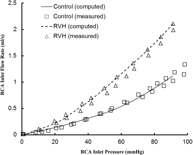
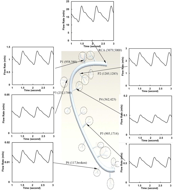
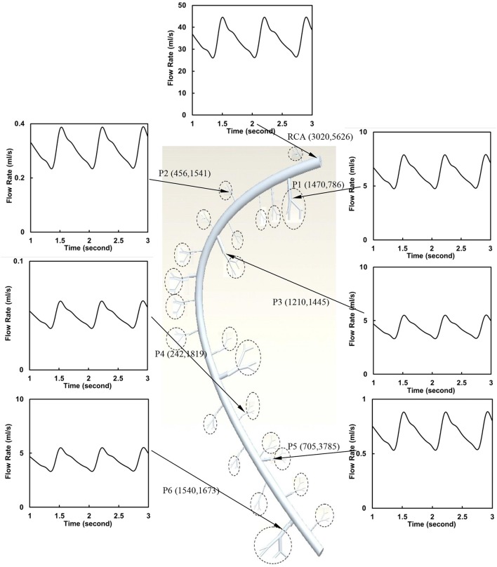
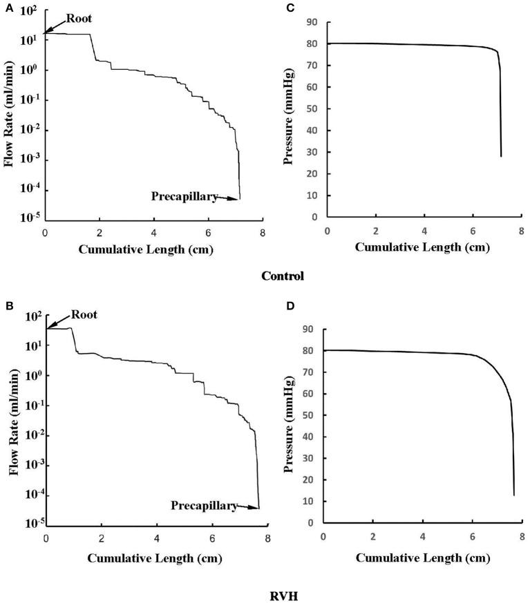
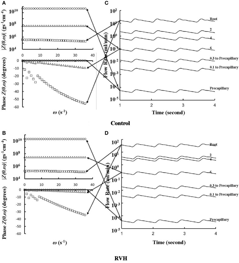
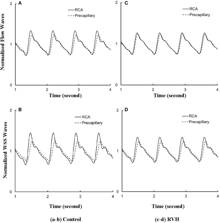
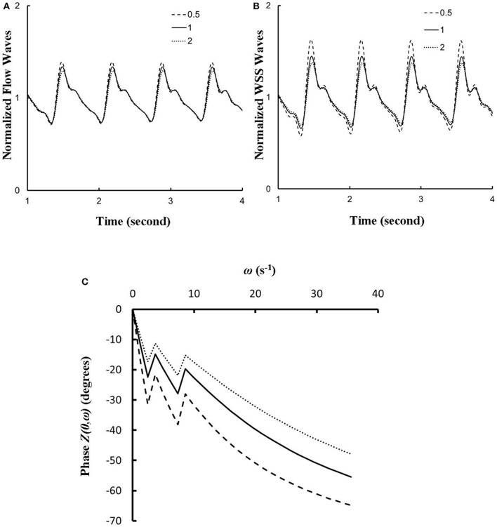
A pulsatile hemodynamic analysis was carried out in the right coronary arterial (RCA) tree of control and RV hypertrophy (RVH) hearts. The shape of flow and wall shear stress (WSS) waves was hypothesized to be maintained throughout the RCA tree in RVH (i.e., similar patterns of normalized flow and WSS waves in vessels of various sizes). Consequently, we reconstructed the entire RCA tree down to the first capillary bifurcation of control and RVH hearts based on measured morphometric data. A Womersley-type model was used to compute the flow and WSS waves in the tree. The hemodynamic parameters obtained from experimental measurements were incorporated into the numerical model. Given an increased number of arterioles, the mean and amplitude of flow waves at the inlet of RCA tree in RVH was found to be two times larger than that in control, but no significant differences (p > 0.05) were found in precapillary arterioles. The increase of stiffness in RCA of RVH preserved the shape of normalized flow and WSS waves, but increased the PWV in coronary arteries and reduced the phase angle difference for the waves between the most proximal RCA and the most distal precapillary arteriole. The study is important for understanding pulsatile coronary blood flow in ventricular hypertrophy.

Keywords: Pulsatile wall shear stress; Womersley-type model; pulsatile flow; right coronary arterial tree; right ventricular hypertrophy.


Figures

Figure 1
Figure 1
Experimental and computed pressure-flow relationship of the RCA of control and RVH hearts. The experimental results were measured under loading and unloading of pressures as the RCA tree was perfused by cardioplegic solution (μ = 1.1 cp and ρ = 1 g/cm3). The pulsatile model was used to compute the pressure and flow at very low frequency (ω → 0).
Figure 2
Figure 2
Flow waves at the inlet and primary branches (P1–P6) of RCA tree of a control heart. The dimensions in the parenthesis refer to diameter and length (μm).
Figure 3
Figure 3
Flow waves at the inlet and primary branches (P1–P6) of RCA tree of a RVH heart. The dimensions in the parenthesis refer to diameter and length (μm).
Figure 4
Figure 4
(A,B) Relationship between the mean flow (averaged over a cardiac cycle) in a vessel and the cumulative length of the vessel from the root to the precapillary arteriole through similar primary branches in: (A) control and (B) RVH pig hearts (i.e., P2 in Figure 2 and P1 in Figure 3, respectively). (C,D) Relationship between the mean pressure (averaged over a cardiac cycle) in a vessel and the cumulative length of the vessel corresponding to (A,B).
Figure 5
Figure 5
(A,B) Impedance |Z(0)| vs. angular frequency ω and phase Z(0) vs. angular frequency ω sequentially along the corresponding paths to Figures 4A,B. (C,D) Flow waves sequentially along the corresponding paths to Figures 4A,B. The arrows indicate the one-to-one correspondence between (A–D).
Figure 6
Figure 6
(A) Flow and (B) WSS waves normalized by the mean values (averaged over a cardiac cycle) at the RCA and precapillary arteriole of control heart. (C,D) Normalized flow and WSS waves at the RCA and precapillary arteriole of RVH heart in correspondence with (A,B), respectively.
Figure 7
Figure 7
(A,B) A sensitivity analysis of normalized (A) flow and (B) WSS waves at the inlet of RCA tree with a 50% increase/decrease of Young's modulus of each vessel wall, where the baseline (i.e., the unity) refers to the static Young's modulus of 8 × 10⁶ dynes/cm2 with adjustment for frequency. (C) Phase Z(0) vs. angular frequency ω at the inlet of RCA tree with a 50% increase/decrease of Young's modulus of each vessel wall.

Similar articles

References

    1. Ashikawa K., Kanatsuka H., Suzuki T., Takishima T. (1986). Phasic blood flow velocity pattern in epimyocardial microvessels in the beating canine left ventricle. Circ. Res. 59, 704–711. 10.1161/01.RES.59.6.704 - DOI - PubMed
    1. Bergel D. H. (1961). The dynamic elastic properties of the arterial wall. J. Physiol. (Lond). 156, 458–469. 10.1113/jphysiol.1961.sp006687 - DOI - PMC - PubMed
    1. Botham M. J., Lemmer J. H., Gerren R. A., Long R. W., Behrendt D. M., Gallagher K. P. (1984). Coronary vasodilator reserve in young dogs with moderate right ventricular hypertrophy. Ann. Thorac. Surg. 38, 101–107. 10.1016/S0003-4975(10)62214-X - DOI - PubMed
    1. Butlin M., Qasem A., Battista F., Bozec E., McEniery C. M., Millet-Amaury E., et al. . (2013). Carotid-femoral pulse wave velocity assessment using novel cuff-based techniques: comparison with tonometric measurement. J Hypertens. 31, 2237–2243, discussion 2243. 10.1097/HJH.0b013e328363c789 - DOI - PubMed
    1. Chiu J. J., Chien S. (2011). Effects of disturbed flow on vascular endothelium: pathophysiological basis and clinical perspectives. Physiol. Rev. 91, 327–387. 10.1152/physrev.00047.2009 - DOI - PMC - PubMed