

Spontaneous rectus sheath hematoma: The utility of CT angiography
- PMID: 29904466
- PMCID: PMC6000050
- DOI: 10.1016/j.radcr.2018.01.016
Spontaneous rectus sheath hematoma: The utility of CT angiography
Abstract
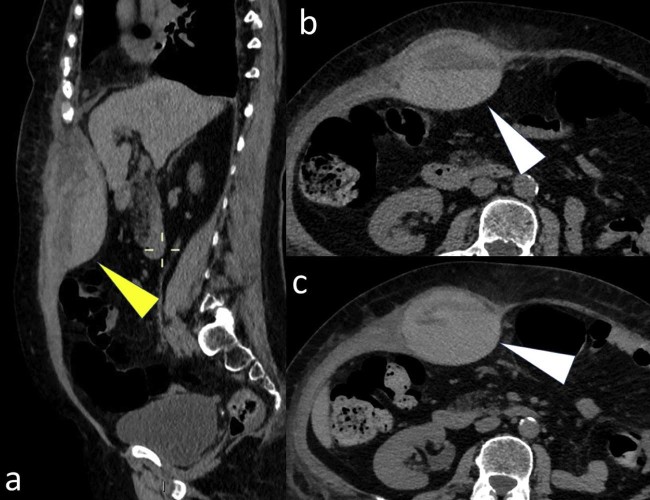
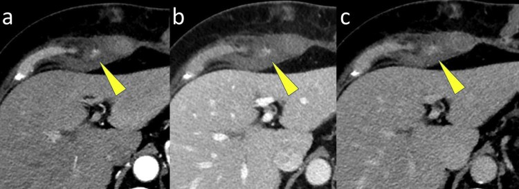
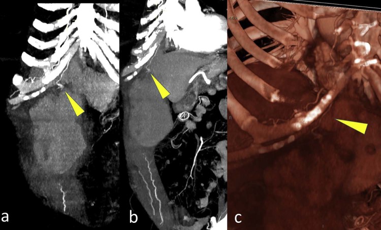
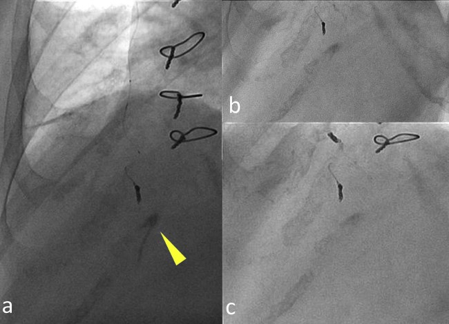
We described the utility of computed tomography (CT) angiography in detection of bleeding vessels for a rapid percutaneous arterial embolization of the spontaneous rectus sheath hematoma. A 70-year-old woman comes to our attention with acute abdominal pain and a low hemoglobin level. An unenhanced CT was performed demonstrating a large rectus sheath hematoma. A conservative management was initially established. Despite this therapy, the abdominal pain increased together with a further decrease of hemoglobin values. A CT angiography was then performed, demonstrating an active bleeding within the hematoma and addressing the patient to a rapid percutaneous arterial embolization.
Keywords: Active bleeding; Acute abdomen; Arterial embolization; CT angiography; Rectus sheath hematoma.
Figures
References
-
- Hatjipetrou A., Anyfantakis D., Kastanakis M. Rectus sheath hematoma: a review of the literature. Int J Surg. 2015;13:267–271. - PubMed
-
- Fitzgerald J.E., Fitzgerald L.A., Anderson F.E., Acheson A.G. The changing nature of rectus sheath haematoma: case series and literature review. Int J Surg. 2009;7(2):150–154. - PubMed
-
- Cherry W.B., Mueller P.S. Rectus sheath hematoma: review of 126 cases at a single institution. Medicine (Baltimore) 2006;85(2):105–110. - PubMed
-
- Berná J.D., Zuazu I., Madrigal M., García-Medina V., Fernández C., Guirado F. Conservative treatment of large rectus sheath hematoma in patients undergoing anticoagulant therapy. Abdom Imaging. 2000;25(3):230–234. - PubMed
-
- Salemis N.S., Gourgiotis S., Karalis G. Diagnostic evaluation and management of patients with rectus sheath hematoma. A retrospective study. Int J Surg. 2010;8(4):290–293. - PubMed
Publication types
LinkOut - more resources
Full Text Sources
Other Literature Sources
