

Case Reports
doi: 10.1016/j.radcr.2018.01.001.
eCollection 2018 Apr.
Variant May-Thurner syndrome: Compression of the left common iliac vein by the ipsilateral internal iliac artery
Affiliations
- PMID: 29904487
- PMCID: PMC5999880
- DOI: 10.1016/j.radcr.2018.01.001
Item in Clipboard
Case Reports
Variant May-Thurner syndrome: Compression of the left common iliac vein by the ipsilateral internal iliac artery
Radiol Case Rep.
.
Abstract
May Thurner syndrome (MTS) is an anatomic variant that can present as acute or chronic deep vein thrombosis. Although it is classically reported in young and middle-aged women, it is also seen in both young and older men. Multiple cases of anatomic variations of MTS have been described. We present an uncommon case of variant MTS, including diagnostic imaging and approach to treatment.
Keywords: Deep vein thrombosis; May-Thurner syndrome; Thrombolysis.
Figures
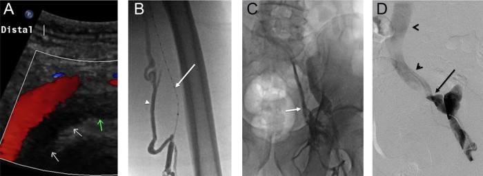
(A) Left lower extremity venous duplex ultrasound demonstrating occlusive deep vein thrombus (DVT) in the external iliac vein (arrows). (B) Extensive left lower extremity occlusive DVT was noted on venography. Note the lysis catheter in the occluded superficial femoral vein (arrow) and filling of collateral muscular veins (arrowhead). (C) Venography demonstrates filling of collaterals draining into the internal iliac vein (arrow). Thrombolysis was initiated in the affected segments. (D) Pelvic venography demonstrated an abrupt change in contrast opacity at the confluence of the external iliac vein with the internal iliac vein (arrow), with the common iliac vein and inferior vena cava remaining patent (arrowheads).
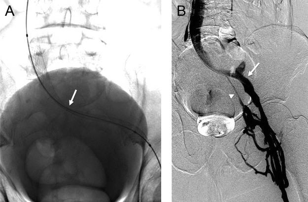
(A) After thrombolysis, balloon angioplasty and stenting was performed, with a significant residual waist at the region of stenosis (arrow). (B) Flow had significantly improved, however, after stenting. Note increased opacification of the external iliac vein (arrow) and less collateral filling of the internal iliac vein (arrowhead).
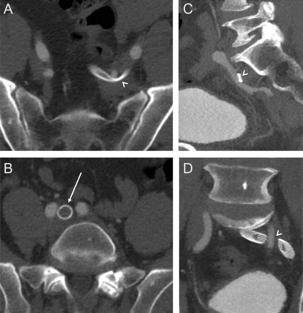
(A, C, D) Computed tomography (CT) with contrast demonstrates compression of the stented iliac veins by the overlying left internal iliac artery (arrowheads). No compression was seen at the confluence of the left common iliac vein with the inferior vena cava (IVC), at the location of typical May-Thurner compression.
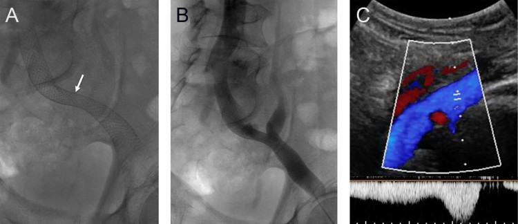
(A) Follow-up radiograph demonstrates interval spontaneous dilation of the waist within the stented left iliac veins (arrow). (B) Venogram demonstrates brisk flow through the stented iliac system. (C) Ultrasound evaluation at follow-up demonstrated a widely patent iliac venous system with normal augmentation of flow.
References
-
- Galang L. May Thurner Syndrome: an important differential diagnosis for DVT. J Vasc Med Surg. 2016;4(2):261.
-
- Kibbe M.R., Ujiki M., Goodwin A.L., Eskandari M., Yao J., Matsumura J. Iliac vein compression in an asymptomatic patient population. J Vasc Surg. 2004;39(5):937–943. - PubMed
-
- Fretz V., Binkert C.A. Compression of the inferior vena cava by the right iliac artery: a rare variant of May-Thurner syndrome. Cardiovasc Intervent Radiol. 2010;33(5):1060–1063. - PubMed
-
- Molloy S., Jacob S., Buckenham T., Khaw K.T., Taylor R.S. Arterial compression of the right common iliac vein; an unusual anatomical variant. Cardiovasc Surg. 2002;10(3):291–292. - PubMed
-
- Steinberg J.B., Jacocks M.A. May-Thurner syndrome: a previously unreported variant. Ann Vasc Surg. 1993;7(6):577–581. - PubMed
Publication types
LinkOut - more resources
Full Text Sources
Other Literature Sources
