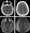
Arachnoid cysts with spontaneous intracystic hemorrhage and associated subdural hematoma: Report of management and follow-up of 2 cases
- PMID: 29904503
- PMCID: PMC5999852
- DOI: 10.1016/j.radcr.2017.12.006
Arachnoid cysts with spontaneous intracystic hemorrhage and associated subdural hematoma: Report of management and follow-up of 2 cases
Abstract
Arachnoid cysts are one of the most frequently encountered intracranial space-occupying lesions in daily neurosurgery and neuroradiology practice. Majority of arachnoid cysts, particularly those of smaller sizes, have a benign uneventful lifetime course. Certain symptoms may indicate serious complications related to underlying arachnoid cysts. Hemorrhage is one of the most fearsome complications of arachnoid cysts and almost all reported cases in the literature have undergone surgical correction. In this study, we aimed to present clinical and radiologic follow-up findings in two adult cases of intracranial arachnoid cyst with spontaneous intracystic hemorrhage and associated subdural hematoma, one of which was successfully treated conservatively. In addition, we broadly summarized and discussed pertinent studies in the English literature.
Keywords: Arachnoid cyst; Headache; Intracystic hemorrhage; Subdural hematoma.
Figures


References
-
- Chen Y., Fang H.J., Li Z.F., Yu S.Y., Li C.Z., Wu Z.B. Treatment of middle cranial fossa arachnoid cysts: a systematic review and meta-analysis. World Neurosurg. 2016;92:480–490. - PubMed
-
- Ildan F., Cetinalp E., Bağdatoğlu H., Boyar B., Uzuneyüoglu Z. Arachnoid cyst with traumatic intracystic hemorrhage unassociated with subdural hematoma. Neurosurg Rev. 1994;17:229–232. - PubMed
-
- Liu Z.1., Xu P., Li Q., Liu H., Chen N., Xu J. Arachnoid cysts with subdural hematoma or intracystic hemorrhage in children. Pediatr Emerg Care. 2014;30:345–351. - PubMed
-
- Kwak Y.S., Hwang S.K., Park S.H., Park J.Y. Chronic subdural hematoma associated with the middle fossa arachnoid cyst: pathogenesis and review of its management. Childs Nerv Syst. 2013;29:77–82. - PubMed
-
- Iaconetta G., Esposito M., Maiuri F., Cappabianca P. Arachnoid cyst with intracystic haemorrhage and subdural haematoma: case report and literature review. Neurol Sci. 2006;26:451–455. - PubMed
Publication types
LinkOut - more resources
Full Text Sources
Other Literature Sources

