

doi: 10.1016/j.vgie.2017.09.004.
eCollection 2018 Jan.
EUS imaging of dead Ascaris
Affiliations
- PMID: 29905174
- PMCID: PMC5965713
- DOI: 10.1016/j.vgie.2017.09.004
Item in Clipboard
EUS imaging of dead Ascaris
VideoGIE.
.
No abstract available
Keywords: CBD, common bile duct.
Figures
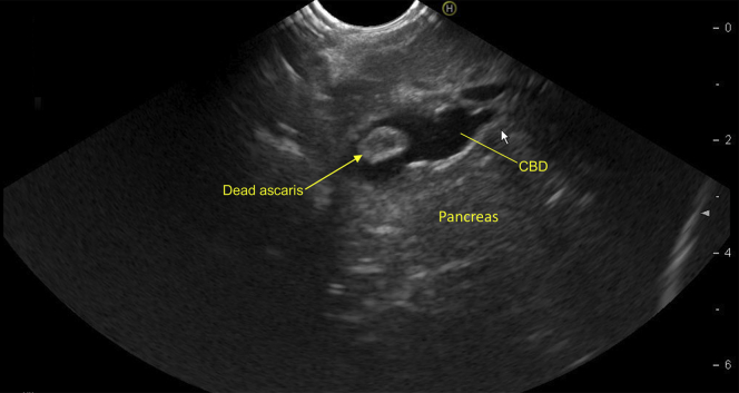
Linear EUS view from the stomach showing dilated CBD with rounded hyperechoic structure without acoustic shadowing in the lower CBD. CBD, common bile duct.

Linear EUS view showing rounded hyperechoic structure in dilated common bile duct. CBD, common bile duct.
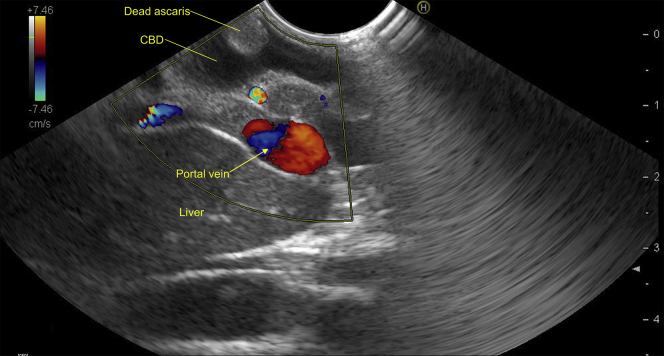
Linear EUS view from duodenal bulb with color Doppler image showing hyperechoic avascular structure with a central anechoic area inside the common bile duct. CBD, common bile duct.

Dual-color Doppler image showing avascular hyperechoic structure with vascular portal vein. CBD, common bile duct.

Zoom image showing dead Ascaris attached to the wall of the CBD from 1 discrete attachment point, whereas the remaining structure was noted to move with the pulsatile flow of biliary secretions. CBD, common bile duct.

The central part of the hyperechoic structure was anechoic. CBD, common bile duct.
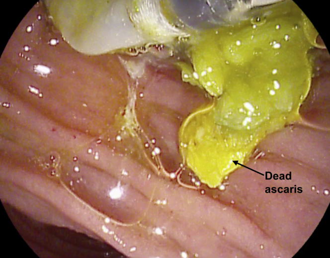
After biliary sphincterotomy and multiple balloon sweeps on ERCP, yellow fragments were removed, suggestive of recently fragmented Ascaris.
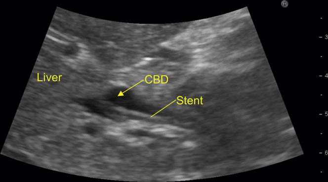
US view of the abdomen showing CBD with stent inside. CBD, common bile duct.
LinkOut - more resources
Full Text Sources
Other Literature Sources
