Review
. 2018 Mar;97(S 01):S48-S113.
doi: 10.1055/s-0043-121596. Epub 2018 Mar 22.

HPV - A different view on Head and Neck Cancer

[Article in English, German]
Affiliations
Review

HPV - A different view on Head and Neck Cancer

[Article in English, German]
Claus Wittekindt et al. Laryngorhinootologie. 2018 Mar.

Abstract

Head and neck cancer is the sixth most common cancer with over 500000 annually reported incident cases worldwide. Besides major risk factors tobacco and alcohol, oropharyngeal squamous cell carcinomas (OSCC) show increased association with human papillomavirus (HPV). HPV-associated and HPV-negative OSCC are 2 different entities regarding biological characteristics, therapeutic response, and patient prognosis. In HPV OSCC, viral oncoprotein activity, as well as genetic (mutations and chromosomal aberrations) and epigenetic alterations plays a key role during carcinogenesis. Based on improved treatment response, the introduction of therapy de-intensification and targeted therapy is discussed for patients with HPV OSCC. A promising targeted therapy concept is immunotherapy. The use of checkpoint inhibitors (e.g. anti-PD1) is currently investigated. By means of liquid biopsies, biomarkers such as viral DNA or tumor mutations in the will soon be available for disease monitoring, as well as detection of treatment failure. By now, primary prophylaxis of HPV OSCC can be achieved by vaccination of girls and boys.

Kopf-Hals-Tumore sind die sechst-häufigste Krebsart mit über 500000 jährlich gemeldeten Fällen weltweit. Die Hauptrisikofaktoren sind Tabak- und Alkoholkonsum, wobei v. a. Oropharynxkarzinome (OSCC) vermehrt eine Assoziation mit humanen Papillomaviren (HPV) aufweisen. Bei HPV-assoziierten und HPV-negativen OSCC handelt es sich bezüglich biologischer Charakteristika, Therapieansprechen und Prognose der Patienten um 2 eigenständige Entitäten, die allerdings bisher identisch behandelt werden. Bei HPV OSCC spielen neben der Aktivität viraler Onkoproteine auch genetische (Mutationen und chromosomale Aberrationen) und epigenetische Veränderungen eine entscheidende Rolle bei der Krebsentstehung. Aufgrund des besseren Therapieansprechens wird aktuell über die Einführung einer De-Intensivierung der Therapie und über zielgerichtete Therapieoptionen für Patienten mit HPV OSCC diskutiert. Ein vielversprechendes zielgerichtetes Therapiekonzept ist bspw. die Immuntherapie. Besonders intensiv wird derzeit die Anwendung von Checkpoint-Inhibitoren (z. B. gegen PD1) erforscht. Mithilfe sogenannter Flüssigbiopsien sollen zukünftig weitere Biomarker, in Form von viraler DNA oder Tumor-Mutationen, zur Überwachung des Krankheitsverlaufs und frühzeitigen Erkennens von Therapieversagen eingesetzt werden. Zur primären Prophylaxe einer Tumorentstehung ist die HPV-Impfung von männlichen und weiblichen Jugendlichen empfehlenswert.


Conflict of interest statement

Die Autoren geben an, dass kein Interessenkonflikt besteht.

Figures

Fig. 1
Fig. 1
Prevalence of oncogenic HPV in OSCC patients who were treated in Giessen is increasing. Currently the prevalence amounts to more than 50%. The data points represent the mean value of 4 years each.
Fig. 2
Fig. 2
The molecular mechanisms of carcinogenesis of HPV- and noxae-associated OSCC (simplified). Dysfunction of the same cellular programs (apoptosis, cell cycle, senescence, and immune system) leads to carcinogenesis in both groups. Multiple genetic mutations that may affect numerous components of the signaling pathways, lead to activation of oncogenes in noxae-associated OSCC and inactivation of tumor suppressors. In contrast, the HPV oncoproteins E5, E6, and E7 lead to interventions in the signaling pathways which dysregulate the same cellular programs. Characteristic for noxae-associated OSCC are mutations of TP53 resulting in inactive p53 overexpression, as well as mutations in the genes coding for Rb and p16INK4A (p16) so that both proteins are reduced. Generally, those mutations are not found in HPV-associated OSCC, and due to the activity of E7, p16 is overexpressed.
Fig. 3
Fig. 3
Frequently, in solid tumors an activation of the hypoxia signaling pathway is found that is obvious due to the central expression of respective marker proteins (here: Glut I) in tumor nests and that can be confirmed by immunohistochemistry (hypoxia-related). Some tumors, however, show a consistently high expression of the same marker that suggest other activation mechanisms of the signaling pathway (endogen-related). The central regulator protein of the hypoxia signaling pathway HIF-1α is present in HPV associated tumor cells in an overexpressed way compared to HPV-negative tumor cells (Western-blot, bottom left). In analogy to fig. 2 , the viral activity leads to activation of the hypoxia signaling pathways and thus to processes that are favorable for carcinogenesis.
Fig. 4
Fig. 4
Immunohistochemical proof of the expression of β2 microglobulin (β2M) as marker of functional membrane-based HLA I expression (top left). The loss of the expression of β2M on tumor cells (bottom left) correlates with a better overall survival of patients with OSCC (right).
Fig. 5
Fig. 5
Immunohistochemical proof of p16INK4A protein expression in single cells of healthy squamous epithelium (top left). Generally, p16INK4A is missing in HPV-negative squamous cell carcinomas of the oropharynx (bottom left). However, strongly overexpressed p16INK4A is present in HPV-associated OSCC (bottom, in the middle) and dysplasia (top right). Single OSCC sometimes show a weak expression of p16INK4A (bottom right) that cannot be considered as positive in the context of HPV-diagnostics.
Fig. 6
Fig. 6
Between 2000 and 2015, the average prevalence of HPV-associated OSCC (HR-HPV DNA and p16INK4A-positive samples) amounted to 23% in Giessen. About 6% of all cases revealed discordant results of HPV DNA and p16INK4A tests, each. The survival of those patients (blue and yellow lines) was significantly inferior compared to patients with HPV-associated OSCC. However, it hardly differed from the survival of patients with HPV-negative OSCC (red line).
Fig. 7
Fig. 7
The current staging for HPV-positive and HPV-negative OSCC was changed significantly. On the left, metastatic HPV OSCC (ipsilateral, <6 cm) is displayed, this means tumor stage I; according to the former edition, the tumor would have to be classified as stage IVa (T1, N2b). On the right, a T3N1 OSCC, HPV-negative, is displayed, to be classified as stage III. Thus, the tumor stage of the patient on the left is lower in comparison to the patient on the right.
Fig. 8
Fig. 8
Because of the new staging rules for HPV OSCC, stage IV tumors are meanwhile rare, so that there is now a relevant percentage of stage I and II tumors.
Fig. 9
Fig. 9
The type of treatment failure in cases of HPV-associated and HPV-negative OSCC is different: patients with HPV-induced OSCC frequently develop distant metastases, in cases of HPV-negative tumors, local and locoregional tumor recurrences prevail.
Fig. 10
Fig. 10
Diagram of the principle of non-invasive liquid biopsy. The circulating tumor cells (CTC), tumor DNA, tumor DNA with integrated HPV DNA as well as episomal HPV DNA are displayed.
Fig. 11
Fig. 11
Correlation between the detection of HPV DNA in blood under therapy as well as during follow-up.
Fig. 12
Fig. 12
Treatment failure in 520 patients with HPV-associated and HPV-negative OSCC. When tumor control fails in HPV-induced tumors, it occurs frequently within the first year after the end of therapy.
Abb. 1
Abb. 1
Die Prävalenz onkogener HPV bei OSCC Patienten, die in Gießen behandelt wurden ist ansteigend. Aktuell liegt die Prävalenz über 50%. Datenpunkte repräsentieren Mittelwerte aus jeweils 4 Jahren.
Abb. 2
Abb. 2
Die molekularen Mechanismen der Karzinogene bei HPV- und Noxen-assoziierten OSCC (vereinfacht). Dysfunktion der gleichen zellulären Programme (Apoptose, Zellzyklus, Seneszenz und Immunsystem) führt in beiden Gruppen zur Karzinogenese. Multiple genetische Veränderungen, die eine Reihe von Komponenten der Signalwege betreffen können, führen bei Noxen-assoziierten OSCC zur Aktivierung von Onkogenen und Inaktivierung von Tumorsuppressoren. Dagegen erfolgen durch die HPV-Onkoproteine E5, E6 und E7 gerichtete Eingriffe in Signalwege, wodurch die gleichen zellulären Programme fehlreguliert werden. Charakteristisch sind bei Noxen-assoziierten OSCC Mutationen in TP53 wodurch häufig inaktives p53 überexprimiert vorliegt, sowie Mutationen in den Genen von Rb und p16INK4A (p16) wodurch beide Proteine reduziert nachweisbar sind. Diese Mutationen fehlen in der Regel bei HPV-assoziierten OSCC und aufgrund der Aktivität von E7 liegt p16 überexprimiert vor.
Abb. 3
Abb. 3
Häufig liegt in soliden Tumoren eine Aktivierung des Hypoxie-Signalwegs vor, die sich in der Expression entsprechender Markerproteinen (hier Glut I) zentral in Tumornestern äußert und immunohistochemisch nachweisbar ist („Hypoxie-bedingt“). Manche Tumore zeigen jedoch eine einheitliche starke Expression der gleichen Marker, die auf andere Aktivierungsmechanismen des Signalwegs schließen lassen („Endogen-bedingt“). Das zentrale Regulatorprotein des Hypoxie-Signalwegs (HIF-1α) liegt bei HPV-assoziierten Tumorzellen im Vergleich zu HPV-negativen Tumorzellen überexprimiert vor (Western-Blot links unten). Analog zu Abb. 2 führt die virale Aktivität zur Aktivierung des Hypoxie-Signalwegs und somit zu Prozessen, die für die Karzinogenese förderlich sind.
Abb. 4
Abb. 4
Immunohistochemischer Nachweis der Expression von β2-Mikroglobulin (β2M) als Marker einer funktionellen, membranständigen HLA I Expression (oben links) . Der Verlust der Expression von β2M auf Tumorzellen (links unten) korreliert mit besserem Gesamtüberleben von Patienten mit OSCC (rechts) .
Abb. 5
Abb. 5
Immunhistochemischer Nachweis der p16INK4A Proteinexpression in einzelnen Zellen von gesundem Plattenepithel (links oben). In der Regel fehlt p16INK4A in HPV-negativen Plattenepithelkarzinomen des Oropharynx (links unten). p16INK4A liegt jedoch bei HPV-assoziierten OSCC (unten Mitte) und Dysplasien (oben rechts) stark überexprimiert vor. Einzelne OSCC zeigen stellenweise eine schwache Expression von p16INK4A (rechts unten), die im Rahmen der HPV-Diagnostik jedoch nicht als positiv zu bewerten ist.
Abb. 6
Abb. 6
Im Zeitraum 2000-2015 beträgt die durchschnittliche Prävalenz HPV-assoziierter OSCC (HR-HPV DNA und p16INK4A positive Proben) in Gießen 23%. Jeweils etwa 6% aller Fälle zeigen diskordante Ergebnisse bei HPV-DNA und p16INK4A Tests. Das Überleben dieser Patienten (blaue und gelbe Kurve) ist signifikant schlechter als das der Patienten mit HPV-assoziierten OSCC, es unterscheidet sich jedoch kaum vom Überleben der Patienten mit HPV-negativen OSCC (rote Kurve).
Abb. 7
Abb. 7
Die aktuelle Stadiengruppierung für HPV positive und HPV negative OSCC wurde umfangreich verändert. Links ist ein metastasiertes HPV OSCC (ipsilateral,<6cm), somit besteht ein Tumorstadium I, nach der alten Auflage wäre der Tumor als Stadium IVa (T1, N2b) zu klassifizieren gewesen. Rechts ist ein T3N1 OSCC, HPV negativ abgebildet, somit als Tumorstadium III zu klassifizieren. Anstatt größer ist das Tumorstadium für den Patienten links nun kleiner im Vergleich zum Patienten rechts.
Abb. 8
Abb. 8
Durch die neuen Stagingregeln für HPV OSCC sind Stadium IV-Tumoren selten geworden, dafür existiert nun ein erheblicher Anteil an Stadium I- und Stadium II-Tumoren.
Abb. 9
Abb. 9
Die Art des Therapieversagens bei HPV-assoziiertem und HPV-negativem OSCC unterscheidet sich: Patienten mit HPV getriebenem OSCC entwickeln häufig Fernemetastasen, bei HPV-negativem Tumor überwiegen lokale oder lokoregionäre Tumorrezidive.
Abb. 10
Abb. 10
Schematische Darstellung des Prinzips einer nicht-invasiven Flüssigbiopsie. Dargestellt sind zirkulierende Tumorzellen (CTC) gesunde zell-freie DNA, Tumor-DNA, Tumor-DNA mit integrierter HPV-DNA sowie episomal vorliegende HPV-DNA.
Abb. 11
Abb. 11
Korrelation zwischen der Detektion von HPV DNA im Blut unter der Therapie sowie während der Nachsorge.
Abb. 12
Abb. 12
Therapieversagen bei 520 Patienten mit HPV-assoziierten und HPV-negativen OSCC. Wenn bei einem HPV-getriebenen Tumor die Tumorkontrolle misslingt, dann häufig frühzeitig innerhalb des ersten Jahres nach Therapieende.

Similar articles

Cited by

References

    1. Javadi P et al.Evolving disparities in the epidemiology of oral cavity and oropharyngeal cancers. Cancer Causes Control. 2017;28:635–645. - PubMed
    1. Mifsud M et al.Evolving trends in head and neck cancer epidemiology: Ontario, Canada 1993–2010. Head Neck. 2017;39:1770–1778. - PubMed
    1. Chaturvedi A K et al.Human papillomavirus and rising oropharyngeal cancer incidence in the United States. J Clin Oncol. 2011;29:4294–4301. - PMC - PubMed
    1. Nasman A et al.Incidence of human papillomavirus (HPV) positive tonsillar carcinoma in Stockholm, Sweden: an epidemic of viral-induced carcinoma? Int J Cancer. 2009;125:362–366. - PubMed
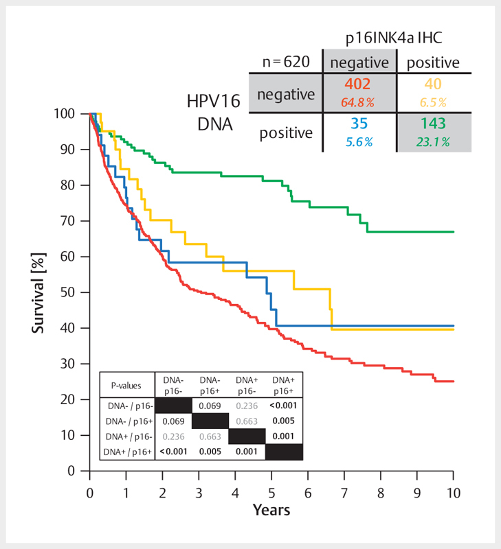
    1. Wittekindt C et al.Expression of p16 protein is associated with human papillomavirus status in tonsillar carcinomas and has implications on survival. Adv Otorhinolaryngol. 2005;62:72–80. - PubMed

MeSH terms