

Radiomics: Big Data Instead of Biopsies in the Future?
- PMID: 29905355
- PMCID: PMC6541032
- DOI: 10.1055/s-0043-121964
Radiomics: Big Data Instead of Biopsies in the Future?
Abstract
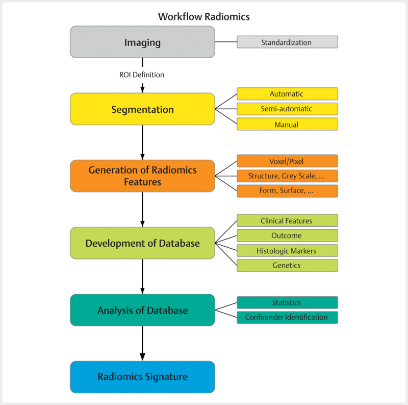
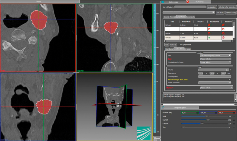
Precision medicine is increasingly pushed forward, also with respect to upcoming new targeted therapies. Individual characterization of diseases on the basis of biomarkers is a prerequisite for this development. So far, biomarkers are characterized clinically, histologically or on a molecular level. The implementation of broad screening methods ("Omics") and the analysis of big data - in addition to single markers - allow to define biomarker signatures. Next to "Genomics", "Proteomics", and "Metabolicis", "Radiomics" gained increasing interest during the last years. Based on radiologic imaging, multiple radiomic markers are extracted with the help of specific algorithms. These are correlated with clinical, (immuno-) histopathological, or genomic data. Underlying structural differences are based on the imaging metadata and are often not visible and therefore not detectable without specific software. Radiomics are depicted numerically or by graphs. The fact that radiomic information can be extracted from routinely performed imaging adds a specific appeal to this method. Radiomics could potentially replace biopsies and additional investigations. Alternatively, radiomics could complement other biomarkers and thus lead to a more precise, multimodal prediction. Until now, radiomics are primarily used to investigate solid tumors. Some promising studies in head and neck cancer have already been published.
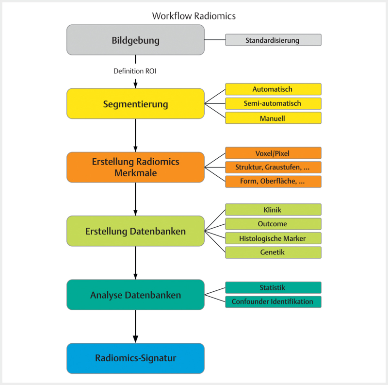
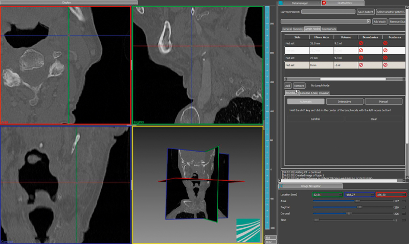
Die Präzisionsmedizin wird – auch mit Zunahme gezielter Therapieoptionen durch Biologicals – immer mehr vorangetrieben. Die individuelle Charakterisierung einer Erkrankung auf der Grundlage von Biomarkern im weitesten Sinne ist eine grundlegende Voraussetzung hierfür. Diese Biomarker können bisher klinisch, histologisch oder molekular bestimmt werden. Die Entwicklung breiter Screening Methoden und gleichzeitig die Möglichkeit, große Datenmengen („Omics“ ) mit geeigneter Software immer besser zu analysieren, führen dazu, dass man sich nicht nur auf einzelne Biomarker beschränkt, sondern Biomarker-Signaturen darstellen kann. Die „Radiomics” finden neben „Genomics“, „Proteomics“ oder „Metabolomics” in den letzten Jahren zunehmendes Interesse und erweitern das Biomarkerfeld. Basierend auf radiologischen Bildern werden eine Vielzahl von Merkmalen mithilfe spezifischer Algorithmen extrahiert. Diese Merkmale werden mit klinischen, (immun-) histopathologischen und genomischen Daten korreliert. Erfasste Strukturunterschiede sind Bestandteil der den Bildern zugrundeliegenden unbearbeiteten Metadaten. Diese sind für das bloße Auge oft nicht sichtbar und insofern ohne spezifische Software für den Untersucher nicht zu erfassen. Sie lassen sich aber numerisch abbilden und grafisch visualisieren. Ein besonderer Reiz der „Radiomics“ ist, dass bereits routinemäßig durchgeführte Bildgebungen einen Biomarker-Charakter zeigen. Dieser hat das Potenzial, Zusatzuntersuchungen oder Biopsien idealerweise durch eine „Radiomics“ Analyse zu ersetzen. Alternativ könnten „Radiomics“-Signaturen andere Biomarker suffizient ergänzen und hierdurch eine präzisere, multimodale Aussage ermöglichen. Bisher finden Radiomics vor allem in der Onkologie solider Tumore Anwendung. Auch bei Kopf-Hals-Karzinomen wurden bereits erste vielversprechende Untersuchungen veröffentlicht.
Eigentümer und Copyright ©Georg Thieme Verlag KG 2018.
Conflict of interest statement
Der Autor gibt an, dass keine Interessenkonflikte bestehen.
Figures

Similar articles
-
Radiomics in Head and Neck Cancer: Extracting Valuable Information from Data beyond Recognition.ORL J Otorhinolaryngol Relat Spec. 2017;79(1-2):65-71. doi: 10.1159/000455704. Epub 2017 Feb 24. ORL J Otorhinolaryngol Relat Spec. 2017. PMID: 28231582 Review.
-
Radiomic biomarkers for head and neck squamous cell carcinoma.Strahlenther Onkol. 2020 Oct;196(10):868-878. doi: 10.1007/s00066-020-01638-4. Epub 2020 Jun 3. Strahlenther Onkol. 2020. PMID: 32495038 Review.
-
[Radiological evaluation of advanced gastric cancer: from image to big data radiomics].Zhonghua Wei Chang Wai Ke Za Zhi. 2018 Oct 25;21(10):1106-1112. Zhonghua Wei Chang Wai Ke Za Zhi. 2018. PMID: 30370508 Review. Chinese.
-
Radiomics: a quantitative imaging biomarker in precision oncology.Nucl Med Commun. 2022 May 1;43(5):483-493. doi: 10.1097/MNM.0000000000001543. Nucl Med Commun. 2022. PMID: 35131965
-
Radiomics and radiogenomics for precision radiotherapy.J Radiat Res. 2018 Mar 1;59(suppl_1):i25-i31. doi: 10.1093/jrr/rrx102. J Radiat Res. 2018. PMID: 29385618 Free PMC article.
Cited by
-
Colorectal liver metastases patients prognostic assessment: prospects and limits of radiomics and radiogenomics.Infect Agent Cancer. 2023 Mar 16;18(1):18. doi: 10.1186/s13027-023-00495-x. Infect Agent Cancer. 2023. PMID: 36927442 Free PMC article. Review.
-
Radiomics and machine learning analysis by computed tomography and magnetic resonance imaging in colorectal liver metastases prognostic assessment.Radiol Med. 2023 Nov;128(11):1310-1332. doi: 10.1007/s11547-023-01710-w. Epub 2023 Sep 11. Radiol Med. 2023. PMID: 37697033
-
Radiomics in radiology: What the radiologist needs to know about technical aspects and clinical impact.Radiol Med. 2024 Dec;129(12):1751-1765. doi: 10.1007/s11547-024-01904-w. Epub 2024 Oct 30. Radiol Med. 2024. PMID: 39472389 Review.
-
Prognostic Assessment of Gastropancreatic Neuroendocrine Neoplasm: Prospects and Limits of Radiomics.Diagnostics (Basel). 2023 Sep 7;13(18):2877. doi: 10.3390/diagnostics13182877. Diagnostics (Basel). 2023. PMID: 37761243 Free PMC article. Review.
-
Radiomics workflow definition & challenges - German priority program 2177 consensus statement on clinically applied radiomics.Insights Imaging. 2024 Jun 3;15(1):124. doi: 10.1186/s13244-024-01704-w. Insights Imaging. 2024. PMID: 38825600 Free PMC article.
References
-
- Giger M L, Karssemeijer N, Schnabel J A. Breast image analysis for risk assessment, detection, diagnosis, and treatment of cancer. Annu Rev Biomed Eng. 2013;15:327–357. - PubMed
Publication types
MeSH terms
Substances
LinkOut - more resources
Full Text Sources
Other Literature Sources