. 2018 Oct;159(10):2058-2065.
doi: 10.1097/j.pain.0000000000001307.

Dynamic sodium imaging at ultra-high field reveals progression in a preclinical migraine model

Affiliations

Dynamic sodium imaging at ultra-high field reveals progression in a preclinical migraine model

Nastaren Abad et al. Pain. 2018 Oct.

Abstract

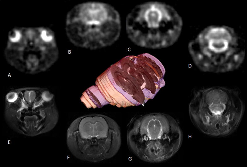
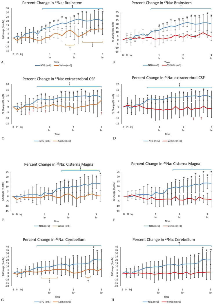
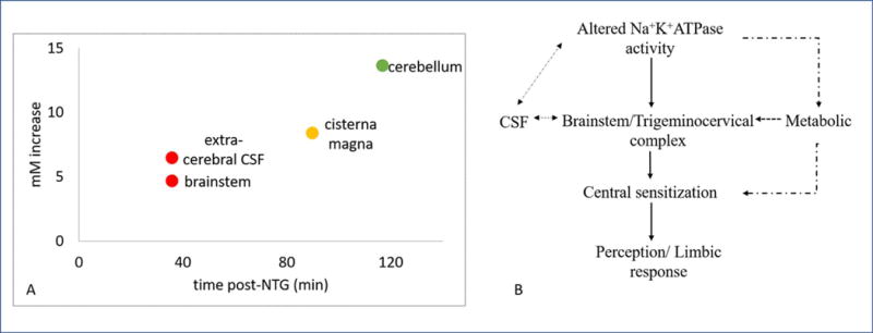
Under the hypothesis that increased extracellular sodium induces sustained neuronal excitability with the onset and progression of migraine, this study evaluates dynamic in vivo Na fluxes in the brain of a preclinical rodent analogue of migraine. Ultra-high field Na magnetic resonance imaging (MRI) at 21.1 T has demonstrated potential to quantify sodium concentrations with good spatial and temporal resolution after the onset of central sensitization. Sprague-Dawley male rats with implanted intraperitoneal lines were studied by MRI before and after an in situ injection of 10 mg/kg of nitroglycerin (NTG) vs vehicle and saline controls. Slice-selective Na images were acquired using a multislice free induction decay-based chemical shift imaging sequence with resolution of 1.1 × 1.1 × 3 mm for a 9-minute acquisition. A total of 27 repeated scans were acquired over 1 hour of baseline scanning and longitudinally up to 3 hours after injection. Increases of Na MRI signal in the brainstem, extracerebral cerebrospinal fluid, and cisterna magna were evident almost immediately after NTG injection, gaining significance from controls in 36 minutes. The cerebellum and third ventricle also showed sustained trends of increased Na, with the former gaining significance at over 2 hours after NTG injection. The data provide evidence of an early change in sodium concentration, markedly in posterior fossa cerebrospinal fluid and brainstem regions. Further study of fluctuations of sodium concentration and their modulation with treatments could help understand the dynamic features of migraine, locate a putative migraine generator, and guide development of therapeutic measures to correct the disturbance of sodium homeostasis.


Conflict of interest statement

Conflict of interest statement

The authors have no conflict of interest to declare.

Figures

Figure 1
Figure 1. Representative sodium and proton MR images displaying relative acquisition locations in the rodent brain
Coronal images (A–D) from a 2D multi-slice 23Na CSI acquired at 1-mm in-plane resolution with a 3-mm slice and (E-H) 117×117-mm 1H multi-slice fast spin-echo images acquired with the same FOV, slice thickness and slice locations. The 3D volume was rendered from a 1H multi-slice data to indicate the 23Na CSI slice locations and thicknesses (blue) relative to the brain (yellow) and a center-slice 1H anatomical reference (grayscale).
Figure 2
Figure 2. Representative coronal images for image segmentation 23Na CSI (top row) with 1H fast spin-echo anatomical references (bottom row)
Regions of interest identified were: (A&E) eyes & olfactory bulb; (B & F) dorsolateral third ventricle, neocortex and muscle; (C & G) extracerebral CSF; (D & H) cisterna magna, brainstem and cerebellum.
Figure 3
Figure 3. Temporal plots for percent change in 23Na concentration
Percent change in 23Na concentration (% change ± SD) for (A-B) brainstem, (C-D) extracerebral CSF, (E-F) cisterna magna and (G-H) cerebellum as a function of time after injection. Signals were normalized to a pre-injection average baseline, with t=0 representative of the pre-injection scan acquired just before the IP injection. Utilizing a mixed model ANOVA, statistical significances are *p<0.05 (Tukey’s post-hoc) for comparisons between (left columns) NTG and saline as control & (right columns) NTG and vehicle as an additional control. For temporal comparisons within the individual cohorts’ statistical significances are identified by †p<0.05 (LSD). (Blue error brackets indicate NTG, whereas orange brackets indicate saline.) Abbreviations: B, Baseline; PI, Pre-Injection; Inj, Injection point; IP, intraperitoneal; SD, Standard Deviation.
Figure 4
Figure 4. Temporally significant sodium increases suggest a hypothesized cascade of neurophysiological events leading to central sensitization
A) Onset of temporal significance for specific brain regions, post administration of NTG to trigger a migraine analogue in rodents. B) Potential neurophysiological cascade of events, elaborated in text, that lead to the onset and progression of NTG triggered acute migraine. The dashed arrows are representative of the hypothesized chain of events that lead to or attenuate the cascade whereas the solid lines are representative of the functional changes taking place.

Similar articles

Cited by

References

    1. Abad N, Rosenberg JT, Roussel T, Grice DC, Harrington MG, Grant SC. Metabolic Assessment of a Migraine Model Using Relaxation-Enhanced 1H Spectroscopy at Ultrahigh Field. Magnetic Resonance in Medicine. 2018;79:1266–1275. - PMC - PubMed
    1. Afridi S, Matharu M, Lee L, Kaube H, Friston K, Frackowiak R, Goadsby P. A PET study exploring the laterality of brainstem activation in migraine using glyceryl trinitrate. Brain. 2005;128:932–939. - PubMed
    1. Akerman S, Holland PR, Goadsby PJ. Diencephalic and brainstem mechanisms in migraine. Nat Rev Neurosci. 2011;12:570–584. - PubMed
    1. Arakaki X, Galbraith G, Pikov V, Fonteh AN, Harrington MG. Altered brainstem auditory evoked potentials in a rat central sensitization model are similar to those in migraine. Brain Res. 2014;1563:110–121. - PMC - PubMed
    1. Ashina M, Hansen JM, Olesen J. Pearls and pitfalls in human pharmacological models of migraine: 30 years’ experience. Cephalalgia. 2013;33:540–553. - PubMed