. 2018 Jun 14;73(7):853-863.
doi: 10.1093/gerona/glx177.

Nrf2 Deficiency Exacerbates Obesity-Induced Oxidative Stress, Neurovascular Dysfunction, Blood-Brain Barrier Disruption, Neuroinflammation, Amyloidogenic Gene Expression, and Cognitive Decline in Mice, Mimicking the Aging Phenotype

Affiliations

Nrf2 Deficiency Exacerbates Obesity-Induced Oxidative Stress, Neurovascular Dysfunction, Blood-Brain Barrier Disruption, Neuroinflammation, Amyloidogenic Gene Expression, and Cognitive Decline in Mice, Mimicking the Aging Phenotype

Stefano Tarantini et al. J Gerontol A Biol Sci Med Sci. .

Abstract

Obesity has deleterious effects on cognitive function in the elderly adults. In mice, aging exacerbates obesity-induced oxidative stress, microvascular dysfunction, blood-brain barrier (BBB) disruption, and neuroinflammation, which compromise cognitive health. However, the specific mechanisms through which aging and obesity interact to remain elusive. Previously, we have shown that Nrf2 signaling plays a critical role in microvascular resilience to obesity and that aging is associated with progressive Nrf2 dysfunction, promoting microvascular impairment. To test the hypothesis that Nrf2 deficiency exacerbates cerebromicrovascular dysfunction induced by obesity Nrf2+/+ and Nrf2-/-, mice were fed an adipogenic high-fat diet (HFD). Nrf2 deficiency significantly exacerbated HFD-induced oxidative stress and cellular senescence, impairment of neurovascular coupling responses, BBB disruption, and microglia activation, mimicking the aging phenotype. Obesity in Nrf2-/- mice elicited complex alterations in the amyloidogenic gene expression profile, including upregulation of amyloid precursor protein. Nrf2 deficiency and obesity additively reduced long-term potentiation in the CA1 area of the hippocampus. Collectively, Nrf2 dysfunction exacerbates the deleterious effects of obesity, compromising cerebromicrovascular and brain health by impairing neurovascular coupling mechanisms, BBB integrity and synaptic function and promoting neuroinflammation. These results support a possible role for age-related Nrf2 dysfunction in the pathogenesis of vascular cognitive impairment and Alzheimer's disease.


Figures
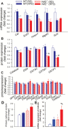
Figure 1.
Figure 1.
Nrf2 deficiency exacerbates obesity-induced oxidative stress. Effects of Nrf2 deficiency and obesity on expression of known Nrf2 target genes at the mRNA (A; quantitative polymerase chain reaction data) and protein (B; quantitative proteomics/selective reaction monitoring [SRM]) level. (C) Effects of Nrf2 deficiency and obesity on protein expression of antioxidant enzymes, whose promoter is not under the control of the Nrf2/ARE system (quantitative proteomics/SRM). (D) Nrf2 deficiency exacerbates high-fat diet (HFD)-induced oxidative stress. Note the significant increases in protein carbonyl content (a marker of oxidative stress) in brain samples from HFD-fed Nrf2-/- mice, which associates with up-regulation of Cdkn2a (p16INK4A; panel E), a marker of increased stress-induced cellular senescence. Data are mean ± SEM (n = 5–6 for each data point). *p < .05 vs Wt (SD), #p < .05 vs Wt (HFD), &p < .05 vs Nrf2-/- (SD) (one-way analysis of variance, Tukey’s post hoc test).
Figure 2.
Figure 2.
Nrf2 deficiency exacerbates obesity-induced neurovascular uncoupling. (A) Representative traces of cerebral blood flow (CBF) measured with a laser Doppler probe above the whisker barrel cortex during contralateral whisker stimulation (30 seconds, 5 Hz) in wild type (Wt) and Nrf2-/- mice fed a high-fat diet (HFD) or standard diet (SD). Panel B depicts the summary data of the effect of HFD-induced obesity on CBF responses to whisker-stimulation in Wt and Nrf2-/- mice. (C) Quantitative polymerase chain reaction data showing HFD-induced changes in mRNA expression of genes relevant for the mediation of neurovascular coupling responses by NO, purinergic signaling and eicosanoid gliotransmitters in the cortices of Wt and Nrf2-/- mice. Data are mean ± SEM (n = 5–10 in each group). *p < .05 vs Wt (SD), #p < .05 vs Wt (HFD), &p < .05 vs Nrf2-/- (SD) (one-way analysis of variance, Tukey’s post hoc test).
Figure 3.
Figure 3.
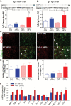
Nrf2 deficiency exacerbates obesity-induced disruption of the blood-brain barrier and promotes neuroinflammation. (A,B) Obesity and Nrf2 deficiency-induced changes in IgG content in the hippocampus of mice. Upper panel: original Western blot showing IgG heavy chain (A) and light chain (B) content in the hippocampi. β-actin was used as a loading control. Bar graphs are summary densitometric values. Data are mean ± SEM. *p < .05 vs Wt (SD), #p < .05 vs Wt (HFD); &p < .05 vs Nrf2-/- (SD). (C) Confocal images showing CD68-positive (red fluorescence, arrowheads) activated microglia in the hippocampi from wild-type SD-fed), wild-type HFD-fed, Nrf2-/- SD-fed, and Nrf2-/- HFD-fed animals. Green fluorescence: Iba-1 microglia marker. Bar graphs are summary data for relative changes in total Iba-1-positive microglia (D) and CD68+/Iba-1+-activated microglia (E). Data are mean ± SEM. *p < .05 vs Wt (SD), #p < .05 vs Wt (HFD); &p < .05 vs Nrf2-/- (SD). (F) Quantitative polymerase chain reaction data showing HFD-induced changes in mRNA expression of proinflammatory cytokines and chemokines and other microglia activation-related factors in the hippocampi of Wt and Nrf2-/- mice. Data are mean ± SEM. *p < .05.
Figure 4.
Figure 4.
(A) Original recordings showing the effects of Nrf2 deficiency and high-fat diet (HFD)-induced obesity on field excitatory postsynaptic potential (EPSP) in the CA1 in response to the stimulation of the Schaffer collaterals on hippocampal brain slices before (10 minutes) and 1 hour after (70 minutes) 4 × 100 Hz tetanus. (B) Long-term potentiation shown as change of field EPSP (fEPSP) slope following a 4 × 100 Hz (1 second) tetanic stimulus in the CA1 of the hippocampus. Data are normalized to baseline responses and depicted as mean ± SEM (n = 8–12 for each data point); *p < .05 vs Wt (SD), #p < .05 vs Wt (HFD) during the last 5 minutes of the recording. (C) The novel object recognition task test used to evaluate recognition memory in mice. The Recognition Index (representing the time spent investigating the novel object relative to the total object investigation) was used as the main index of retention. Data are mean ± SEM. *p < .05 vs Wt (SD). (D) Proposed scheme depicting the likely role of age-related Nrf2 dysfunction in exacerbation of obesity-induced oxidative stress, BBB disruption, neuroinflammation, neurovascular dysfunction, and synaptic dysfunction, all of which contribute to cognitive impairment in obese elderly subjects.

Similar articles

Cited by

References

    1. Wolf PA, Beiser A, Elias MF, Au R, Vasan RS, Seshadri S. Relation of obesity to cognitive function: importance of central obesity and synergistic influence of concomitant hypertension. The Framingham Heart Study. Curr Alzheimer Res. 2007;4:111–116. - PubMed
    1. Gustafson DR, Karlsson C, Skoog I, Rosengren L, Lissner L, Blennow K. Mid-life adiposity factors relate to blood-brain barrier integrity in late life. J Intern Med. 2007;262:643–650. doi:10.1111/j.1365-2796.2007.01869.x - PubMed
    1. Beydoun MA, Beydoun HA, Wang Y. Obesity and central obesity as risk factors for incident dementia and its subtypes: a systematic review and meta-analysis. Obes Rev. 2008;9:204–218. doi:10.1111/j.1467-789X.2008.00473.x - PMC - PubMed
    1. Alosco ML, Spitznagel MB, Raz N et al. . Obesity interacts with cerebral hypoperfusion to exacerbate cognitive impairment in older adults with heart failure. Cerebrovasc Dis Extra. 2012;2:88–98. doi:10.1159/000343222 - PMC - PubMed
    1. Tarantini S, Tran CH, Gordon GR, Ungvari Z, Csiszar A. Impaired neurovascular coupling in aging and Alzheimer’s disease: contribution of astrocyte dysfunction and endothelial impairment to cognitive decline. Exp Gerontol. 2016. doi:10.1016/j.exger.2016.1011.1004 - PMC - PubMed

Publication types

MeSH terms