. 2018 Jul 27;46(13):6746-6760.
doi: 10.1093/nar/gky479.

sRNA-dependent control of curli biosynthesis in Escherichia coli: McaS directs endonucleolytic cleavage of csgD mRNA

Affiliations

sRNA-dependent control of curli biosynthesis in Escherichia coli: McaS directs endonucleolytic cleavage of csgD mRNA

Patrick Rosendahl Andreassen et al. Nucleic Acids Res. .

Abstract

Production of curli, extracellular protein structures important for Escherichia coli biofilm formation, is governed by a highly complex regulatory mechanism that integrates multiple environmental signals through the involvement of numerous proteins and small non-coding RNAs (sRNAs). No less than seven sRNAs (McaS, RprA, GcvB, RydC, RybB, OmrA and OmrB) are known to repress the expression of the curli activator CsgD. Many of the sRNAs repress CsgD production by binding to the csgD mRNA at sites far upstream of the ribosomal binding site. The precise mechanism behind sRNA-mediated regulation of CsgD synthesis is largely unknown. In this study, we identify a conserved A/U-rich region in the csgD mRNA 5' untranslated region, which is cleaved upon binding of the small RNAs, McaS, RprA or GcvB, to sites located more than 30 nucleotides downstream. Mutational analysis shows that the A/U-rich region as well as an adjacent stem-loop structure are required for McaS-stimulated degradation, also serving as a binding platform for the RNA chaperone Hfq. Prevention of McaS-activated cleavage completely relieves repression, suggesting that endoribonucleolytic cleavage of csgD mRNA is the primary regulatory effect exerted by McaS. Moreover, we find that McaS-mediated degradation of the csgD 5' untranslated region requires RNase E.


Figures

Figure 1.
Figure 1.
McaS producing cells lack extracellular curli fibers. Close inspection of the inhibitory effect of McaS on curli fibers. Wild-type cells of SØ928ΔmcaS producing McaS from a Plac containing low-copy plasmid (pNDM-mcaS) and SØ928ΔmcaS cells carrying the empty vector (pNDM220) were grown in curli stimulating conditions on LB-agar plates (without NaCl) at 28°C. High-resolution EM images were taken after 48 hours of growth in the presence of 100 μM IPTG inducer.
Figure 2.
Figure 2.
An A/U-rich stretch followed by a small stem–loop structure is conserved in the csgD mRNA. csgD from seven different species, all having a conserved mcaS gene, was aligned using Clustal Omega Multiple Sequence Alignment tool. (A) The promoter elements (−35 and −10) and transcriptional start site (+1) are highly conserved, as well as the McaS-binding site (dark gray), the RBS and stem–loop II. Furthermore, both an A/U-rich region (light gray) and a C and a G rich stretch (C-side and G-side, respectively) just downstream the A/U-rich region are conserved. Arrows indicate possible stem–loop structures (numbered I, II and III), bold letters indicate nucleotide residues engaging in the base-pairing of the stem–loops, and underlined lower-case letters indicate the translation start codon. (B) The E. coli csgD 5′UTR used in this study adopts an overall similar fold as the consensus folding shown in C. The E. coli structure is based on the structural probing presented in Figure 7 and supplementary data. The two McaS-binding sites are highlighted (C) RNAalifold was used to predict a generic consensus secondary structure of the csgD alignment obtained from Clustal Omega. The consensus sequence predicts csgD to contain three stem–loop structures: the first just downstream of the A/U-rich region (I), a second central large stem–loop structure upstream of the RBS containing a bulge (II), and a third stem–loop structure containing the RBS (III).
Figure 3.
Figure 3.
McaS, RprA, and GcvB stimulate csgD mRNA cleavage in the conserved A/U-rich region. (A) Primer extension and northern blot with RNA purified from SØ928Δ5 carrying pBAD-csgDchiX and either pNDM-mcaS, pNDM-rprA, pNDM-gcvB, pNDM-omrA or the empty vector pNDM220. Cultures were grown in LB broth at 37° to an OD600 of 0.6 (−/−). The cultures were induced with 1 mM IPTG for 10 min (+/−), followed by 5 min induction with 0.2% arabinose (+/+). Primer extension and northern blot was performed as described in methods. (B) csgD mRNA sequence with cleavage sites indicated. The most apparent cleavage sites (formula image) are marked with black arrows and the remaining (formula imageformula imageformula imageformula image) with grey arrows. formula imageis specific to McaS, while formula imageis specific to GcvB. McaS, RprA and GcvB all induce cleavage in the conserved A/U-rich region (gray bar).
Figure 4.
Figure 4.
Only the upstream binding site is necessary for McaS-mediated regulation of CsgD protein levels. (A) Schematic presentation of the csgD 5′UTR indicating the two sites of csgD complementary to McaS (underlined). Deletion mutants in binding sites 1 and 2 respectively are highlighted by black circles. (B) E. coli strain SØ928 carrying either a wild-type csgDFLAG construct or mutant alleles on a pBAD plasmid vector. Expression from the empty vector control pNDM220 (−) or the isogenic plasmid borne McaS (+) was induced by addition of 1 mM IPTG. After 10 min of incubation, 0,2% arabinose was added to induced expression of the csgDFLAGgenes. After 5 min of induction samples for western blot analysis was taken. GroEL was probed as loading control.
Figure 5.
Figure 5.
The A/U-rich region and secondary structure is important for efficient mRNA cleavage and translational inhibition. (A) Graphical representation of wild-type (WT) and mutant csgD mRNAs, shown with consensus secondary structure (Figure 2) indicating the conserved A/U-rich region (grey line), nucleotide substitutions (asterisk) and cleavage site formula imageand formula imagefrom Figure 3 (arrows). (B) Northern blot with RNA purified from SØ928Δ5 carrying either wild-type or mutant pBAD-csgDchiX derivatives (this plasmid contains the csgD 5′UTR and the first 87 nucleotides of the coding region and the small RNA ChiX for stabilisation of the construct) and either pNDM-mcaS or the empty vector pNDM220. Cultures were grown in LB media at 37°C to an OD600 of 0.6 at which point a sample was taken from the culture with wild type csgD and empty pNDM220 vector as a negative control. The cultures were induced with 1 mM IPTG for 10 min, followed by 5 min induction with 0.2% arabinose at which point samples were taken from all cultures. Northern blot was performed as described in methods. (C) Northern and Western blot with RNA and proteins purified from SØ928Δ5 carrying either wild type or mutant pBAD-csgDFLAG and either pNDM-mcaS or the empty vector pNDM220. Cultures were grown and samples were taken as for panel B and Western and northern blots were performed as described in methods. The CsgD-FLAG levels were quantified from three experiments and normalized to the loading control (GroEL). The efficiency of McaS-mediated repression was determined by dividing the signal from samples without McaS by the signal from samples with McaS. Efficiency of McaS on wild type csgD was defined as 1.
Figure 6.
Figure 6.
Hfq binds the csgD 5′UTR constructs with different affinities assisted by the adjacent conserved stem–loop. Electrophoretic mobility shift assays of Hfq binding to csgD mRNA. Samples containing 5′ end-labeled transcripts of 2 nM csgD wild type or csgD mutant mRNAs (Figure 4A) were incubated with increasing amounts of Hfq. Final monomeric concentrations of Hfq were 0, 0.25, 0.5, 0.75, 1 and 2 μM from left to right.
Figure 7.
Figure 7.
Hfq interacts with the csgD 5′UTR at multiple positions. (A) Structural probing assays of csgD 5′ UTR RNA using Nuclease S1 with increasing concentrations of Hfq, McaS or both. Samples containing 5′end-labeled transcripts of 2 nM csgD 5′UTR RNA were pre-incubated at 37°C with Hfq, McaS or both for 10 min before incubation with Nuclease S1 for 5 min. An alkaline hydrolysis ladder (‘OH’) and a G-ladder (‘T1’, RNase T1 digestion) was used to determine nucleotide position. Untreated csgD 5′ UTR RNA was used as a control (‘C’). (B) Protected Hfq and McaS binding sites are underlined in the csgD 5′ UTR structure.
Figure 8.
Figure 8.
McaS mediated csgD mRNA cleavage is RNase E dependent. Western blot of CsgDFLAG and primer extension analysis of csgD 5′end. Cultures of N3431 (rne-3071), the isogenic N3433 WT strain, containing the OmpR234 point mutation and a single or double knock-outs of rng, the gene encoding RNase G. The strains carry either the empty vector control pBAD33 or pBAD-mcaS. All cultures were grown in LB medium at 30°C in the presence of inducer (0.2% arabinose) to an OD600 of 1.2. At this point, the cultures were shifted to 42°C for 20 min before sampling for proteins and RNA. GroEL was probed for protein loading control and 5S, showing characteristic unprocessed bands in the rne-3071 allele, was probed as RNA loading control. Fold-change are of CsgDFLAG protein levels from three experiments.
Figure 9.
Figure 9.
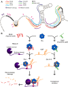
Model for sRNA-mediated regulation of csgD. (A) Binding/cleavage sites of the sRNAs, Hfq and RNase E on the csgD mRNA. McaS, RprA, GcvB and OmRA/B all have extensive base pairing located distally from the ribosomal binding site. While OmrA and OmrB cause translational inhibition, McaS, RprA and GcvB induce ribonucleolytic cleavage of the mRNA. The most significant cleavage site is located near the 5′-end, just upstream of an Hfq binding site. Mutations of this Hfq site inhibits RNase E cleavage and alleviates McaS induced repression of CsgD expression. RydC and RybB bind csgD at the ribosomal binding site to inhibit the formation of the translational initiation complex. (B) Model for the two sRNA mediated mechanisms for posttranscriptional regulation of csgD. Left) McaS (RprA and GcvB) is first bound and stabilized by Hfq. The sRNA-Hfq binary complex interacts with RNase E of the degradosome and its substrate mRNA through base-pairing between the RNAs as well as protein-RNA and protein-protein interactions, in which Hfq binds to Site I of csgD. Complex formation leads to ribonucleolytic cleavage of csgD and McaS. Right) RydC (omrA/B and rybB) is bound by Hfq. This sRNA-Hfq complex will bind near the ribosomal binding site by base-pairing between the two RNAs as well as Hfq interactions with csgD at site II and site III. Formation of this complex leads to translational inhibition.

References

    1. Flemming H.C., Wingender J.. The biofilm matrix. Nat. Rev. Microbiol. 2010; 8:623–633. - PubMed
    1. Hall-Stoodley L., Costerton J.W., Stoodley P.. Bacterial biofilms: from the natural environment to infectious diseases. Nat. Rev. Microbiol. 2004; 2:95–108. - PubMed
    1. Espeland E.M., Wetzel R.G.. Complexation, stabilization, and UV photolysis of extracellular and surface-bound glucosidase and alkaline phosphatase: implications for biofilm microbiota. Microb. Ecol. 2001; 42:572–585. - PubMed
    1. Hoiby N., Ciofu O., Johansen H.K., Song Z.J., Moser C., Jensen P.O., Molin S., Givskov M., Tolker-Nielsen T., Bjarnsholt T.. The clinical impact of bacterial biofilms. Int. J. Oral Sci. 2011; 3:55–65. - PMC - PubMed
    1. White A.P., Gibson D.L., Collinson S.K., Banser P.A., Kay W.W.. Extracellular polysaccharides associated with thin aggregative fimbriae of Salmonella enterica serovar enteritidis. J. Bacteriol. 2003; 185:5398–5407. - PMC - PubMed

Publication types

MeSH terms

LinkOut - more resources