. 2018 Jul 27;46(13):6455-6469.
doi: 10.1093/nar/gky444.

5-Formylcytosine mediated DNA-protein cross-links block DNA replication and induce mutations in human cells

Affiliations

5-Formylcytosine mediated DNA-protein cross-links block DNA replication and induce mutations in human cells

Shaofei Ji et al. Nucleic Acids Res. .

Erratum in

Abstract

5-Formylcytosine (5fC) is an epigenetic DNA modification introduced via TET protein-mediated oxidation of 5-methyl-dC. We recently reported that 5fC form reversible DNA-protein conjugates (DPCs) with histone proteins in living cells (Ji et al. (2017) Angew. Chem. Int. Ed., 56:14130-14134). We now examined the effects of 5fC mediated DPCs on DNA replication. Synthetic DNA duplexes containing site-specific DPCs between 5fC and lysine-containing proteins and peptides were subjected to primer extension experiments in the presence of human translesion synthesis DNA polymerases η and κ. We found that DPCs containing histones H2A or H4 completely inhibited DNA replication, but the replication block was removed when the proteins were subjected to proteolytic digestion. Cross-links to 11-mer or 31-mer peptides were bypassed by both polymerases in an error-prone manner, inducing targeted C→T transitions and -1 deletions. Similar types of mutations were observed when plasmids containing 5fC-peptide cross-links were replicated in human embryonic kidney (HEK) 293T cells. Molecular simulations of the 11-mer peptide-dC cross-links bound to human polymerases η and κ revealed that the peptide fits well on the DNA major groove side, and the modified dC forms a stable mismatch with incoming dATP via wobble base pairing in the polymerase active site.


Figures

Figure 1.
Figure 1.
(A) The formation of DNA–protein and DNA–peptide cross-links at 5-formylcytosine of DNA. The aldehyde group of 5fC readily forms reversible Schiff base conjugates with the Arg or Lys side chains of proteins and polypeptides, which can be stabilized by reduction with NaCNBH3 or glutathione (GSH). (B) Representative MALDI MS spectrum of the conjugate between 5fC (X) containing DNA 23-mer 5′-AGG GTT TTC CXA GTC ACG ACG TT-3′ and 11-mer polypeptide RPKPQQFFGLM-CONH2 following NaCNBH3 reduction. (C) Denaturing PAGE analysis of the cross-links between 5fC (X) containing DNA 23-mer 5′-AGG GTT TTC CXA GTC ACG ACG TT-3′ and histone H2A. The reaction mixtures were heated prior to loading on gel, and samples in lanes 3 and 4 were reduced with NaCNBH3 or GSH, respectively, to stabilize DPCs.
Figure 2.
Figure 2.
Primer extension assays for hPol η and hPol κ catalyzed bypass of reversible DNA–histone cross-links generated via a Schiff base formation between histone proteins and 5fC in DNA. 32P-labeled 12-mer primer was annealed with 23-mer template containing 5-formyl-dC (A), or 5-formyl-dC conjugated to histone H2A via Schiff base formation (B). Polymerase reactions were initiated by addition of a mixture of hPol η or κ and a mixture of dNTP, quenched at pre-selected time points, and loaded onto 20% denaturing PAGE. Percent bypass was plotted over time (C).
Figure 3.
Figure 3.
Primer extension assays for replication bypass of reductively stabilized DNA–protein or DNA–peptide cross-links by hPol η (top panel) and hPol κ (bottom panel). 32P-labeled 12-mer primer was annealed with 23-mer template containing unmodified dC (A), 5-formyl-dC (B), or C5-dC cross-links to 11-mer peptide RPKPQQFFGLM-CONH2 (C) 31-mer peptide YGGFMTSEKSQTPLVTLFKNAIIKNAYKKGE (D), full size histone H2A (E), histone H4 (F), or proteinase K digested histone H4 (G). Polymerase reactions were initiated by addition of DNA polymerases and a mixture of dNTP and quenched at pre-selected time points prior to loading onto 20% denaturing PAGE.
Figure 4.
Figure 4.
Single nucleotide insertion opposite unmodified dC (control) and 5fC-11 mer peptide cross-links (X = RPKPQQFFGLM-CONH2). template–primer complexes (top) were incubated with hPol η (top panel) or hPol κ (bottom panel) in the presence of 50 μM individual dNTP and then quenched at pre-selected time points.
Figure 5.
Figure 5.
HPLC–ESI-MS/MS sequencing of primer extension products via streptavidin capture- nanoHPLC–ESI-MS/MS (A) and MS/MS spectra of primer extension products 5′-pGAC TGG GAA-3′ (B, error free), 5′-pGAC TAG GAA-3′ (C, C → T transition), and 5′-pGAC T_G GAA-3′ (D, –1 deletion).
Figure 6.
Figure 6.
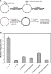
(A) Construction of DPC-containing pMS2 plasmid and its replication in HEK 293T cells. (B)Types and frequencies of mutations induced by the DNA–peptide cross-link in HEK293T cells. The data represent the mean and standard deviation of three independent experiments. Targeted C → T, C → G, C deletion, 3–6 base deletions as well as a variety of semi-targeted mutations near the lesion sites.
Figure 7.
Figure 7.
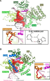
(A) The most representative structure from the 400 ns MD simulation of the pol η-DNA ternary complex, containing the 11-mer peptide cross-linked to the templating C opposite to the N1 protonated incoming dATP. Inset: (left), a zoomed-in view of the 11-mer peptide cross-linked to the templating C opposite the incoming dATP; (right), hydrogen bonding scheme of the C–A+ wobble pair. The protonated hydrogen at N1 in adenine is shown in red. A rotating view is given in the Supplementary Movie (Movie S1). (B) The most representative structure from the 400 ns MD simulation of the pol κ–DNA ternary complex, containing the 11-mer peptide cross-linked to the templating C opposite to the N1 protonated dATP. Inset: a zoomed-in view. A rotating view is given in the Supplementary Movie (Movie S2). In both structures, the complex is displayed in cartoon diagram except for the dATP and the cross-link-containing template C, which are in stick, and Mg2+ ions which are shown as yellow balls. The 11-mer peptide is shown in red surface. The two hydrogen bonds between the template base and incoming nucleotide are shown with dotted lines.

References

    1. Barker S., Weinfeld M., Murray D.. DNA–protein crosslinks: their induction, repair, and biological consequences. Mutat. Res. 2005; 589:111–135. - PubMed
    1. Ide H., Shoulkamy M.I., Nakano T., Miyamoto-Matsubara M., Salem A.M.H.. Repair and biochemical effects of DNA–protein crosslinks. Mutat. Res. 2011; 711:113–122. - PubMed
    1. Tretyakova N.Y., Groehler A., Ji S.. DNA–protein cross-links: formation, structural identities, and biological outcomes. Acc. Chem. Res. 2015; 48:1631–1644. - PMC - PubMed
    1. Barker S., Weinfeld M., Zheng J., Li L., Murray D.. Identification of mammalian proteins cross-linked to DNA by ionizing radiation. J. Biol. Chem. 2005; 280:33826–33838. - PubMed
    1. Speit G., Schütz P., Merk O.. Induction and repair of formaldehyde-induced DNA–protein crosslinks in repair-deficient human cell lines. Mutagenesis. 2000; 15:85–90. - PubMed

Publication types